
ICD-9-CM 95.21 | MeSH D004596 | |
 | ||
Electroretinography measures the electrical responses of various cell types in the retina, including the photoreceptors (rods and cones), inner retinal cells (bipolar and amacrine cells), and the ganglion cells. Electrodes (DTL silver/nylon fiber string) are usually placed on the surface of the cornea for Full Field/Global/Multifocal ERG's and brass/copper electrodes are placed on the skin near the eye for EOG type testing. During a recording, the patient's eyes are exposed to standardized stimuli and the resulting signal is displayed showing the time course of the signal's amplitude (voltage). Signals are very small, and typically are measured in microvolts or nanovolts. The ERG is composed of electrical potentials contributed by different cell types within the retina, and the stimulus conditions (flash or pattern stimulus, whether a background light is present, and the colors of the stimulus and background) can elicit stronger response from certain components.
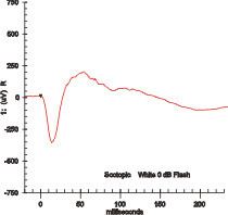
If a dim flash ERG is performed on a dark-adapted eye, the response is primarily from the rod system. Flash ERGs performed on a light adapted eye will reflect the activity of the cone system. Sufficiently bright flashes will elicit ERGs containing an a-wave (initial negative deflection) followed by a b-wave (positive deflection). The leading edge of the a-wave is produced by the photoreceptors, while the remainder of the wave is produced by a mixture of cells including photoreceptors, bipolar, amacrine, and Muller cells or Muller glia. The pattern ERG (PERG), evoked by an alternating checkerboard stimulus, primarily reflects activity of retinal ganglion cells.
Clinically used mainly by ophthalmologists and optometrists, the electroretinogram (ERG) is used for the diagnosis of various retinal diseases.
Inherited retinal degenerations in which the ERG can be useful include:
Other ocular disorders in which the standard ERG provides useful information include:
The ERG is also used extensively in eye research, as it provides information about the function of the retina that is not otherwise available.
Other ERG tests, such as the Photopic Negative Response (PhNR) and pattern ERG (PERG) may be useful in assessing retinal ganglion cell function in diseases like glaucoma.
The multifocal ERG is used to record separate responses for different retinal locations.
The international body concerned with the clinical use and standardization of the ERG, EOG, and VEP is the International Society for the Clinical Electrophysiology of Vision (ISCEV)
In addition to its clinical diagnostic purpose, the ERG can be used during the course of drug development and in clinical trails for testing ocular safety and efficacy of new or existing drugs and treatment modalities