
Dorlands/Elsevier f_05/12360925 | ||
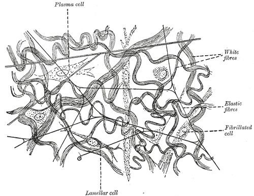 | ||
Elastic fibers (or yellow fibers) are bundles of proteins (elastin) found in extracellular matrix of connective tissue and produced by fibroblasts and smooth muscle cells in arteries. These fibers can stretch up to 1.5 times their length, and snap back to their original length when relaxed. Elastic fibers include elastin, elaunin and oxytalan.
Contents
Elastic tissue is classified as "connective tissue proper".
The elastic fiber is formed from the elastic microfibril (consisting of numerous proteins such as microfibrillar-associated glycoproteins, fibrillin, fibullin, and the elastin receptor) and amorphous elastin.
The microfibril scaffolds and organizes the deposition of amorphous elastin. Amorphous elastin forms from monomers of soluble tropoelastin which is insolubilized and crosslinked into amorphous elastin by lysyl oxidase. Lysyl oxidase reacts with specific lysine residues and by oxidative deamination generates reactive aldehydes and allysine.
These reactive aldehydes and allysines can react with lysine and other allysine residues to crosslink and form desmosine, isodesmosine, and a number of other polyfunctional crosslinks that join surrounding elastin molecules to build an elastin matrix and elastic fiber. These unique crosslinks are responsible for elastin's elasticity.
Distribution
Elastic fibers are found in the skin, lungs, arteries, veins, connective tissue proper, elastic cartilage, periodontal ligament, fetal tissue and other structures. Elastic fibers are absent from scarring, keloids and dermatofibromas and they are decreased greatly, or are absent in anetodermas.
Histology
Elastic fibers stain well with aldehyde fuchsin, orcein, and Weigert's elastic stain in histological sections.
The permanganate-bisulfite-toluidine blue reaction is a highly selective and sensitive method for demonstrating elastic fibers under polarizing optics. The induced birefringence demonstrates the highly ordered molecular structure of the elastin molecules in the elastic fiber. This is not readily apparent under normal optics.
Defects and disease
There is evidence to believe that certain defects of any components of the elastic matrix may impair and alter the structural appearance of elastic and collagen fibers.
Cutis laxa and Williams syndrome have elastic matrix defects that have been directly associated with alterations in the elastin gene.
Alpha-1 antitrypsin deficiency is a genetic disorder where elastin is excessively degraded by elastase, a degrading protein released by neutrophils during the inflammatory response. This leads most often to emphysema and liver disease in affected individuals.
Buschke-Ollendorff syndrome, Menkes disease, pseudoxanthoma elasticum, and Marfan's syndrome have been associated with defects in copper metabolism and lysyl oxidase or defects in the microfibril (defects in fibrillin, or fibullin for example).
Hurler disease, a lysosomal storage disease, is associated with an altered elastic matrix.
Hypertension and some congenital heart defects are associated with alterations in the great arteries, arteries, and arterioles with alterations in the elastic matrix.