
Entrez 2067 | Ensembl ENSG00000012061 | |
 | ||
Aliases ERCC1, COFS4, RAD10, UV20, excision repair cross-complementation group 1, ERCC excision repair 1, endonuclease non-catalytic subunit External IDs MGI: 95412 HomoloGene: 1501 GeneCards: ERCC1 | ||
DNA excision repair protein ERCC-1 is a protein that in humans is encoded by the ERCC1 gene. Together with ERCC4, ERCC1 forms the ERCC1-XPF enzyme complex that participates in DNA repair and DNA recombination.
Contents
- Gene
- Protein
- Structure specific nuclease
- Nucleotide excision repair
- DNA double strand break repair
- DNA interstrand crosslink repair
- Cerebro oculo facio skeletal syndrome
- Cockayne syndrome
- Relevance in chemotherapy
- Deficiency in cancer
- References
Many aspects of these two gene products are described together here because they are partners during DNA repair. The ERCC1-XPF nuclease is an essential activity in the pathway of DNA nucleotide excision repair (NER). The ERCC1-XPF nuclease also functions in pathways to repair double-strand breaks in DNA, and in the repair of “crosslink” damage that harmfully links the two DNA strands.
Cells with disabling mutations in ERCC1 are more sensitive than normal to particular DNA damaging agents, including ultraviolet (UV) radiation and to chemicals that cause crosslinking between DNA strands. Genetically engineered mice with disabling mutations in ERCC1 have defects in DNA repair, accompanied by metabolic stress-induced changes in physiology that result in premature aging. Complete deletion of ERCC1 is incompatible with viability of mice, and no human individuals have been found with complete (homozygous) deletion of ERCC1. Rare individuals in the human population harbor inherited mutations that impair the function of ERCC1. When the normal genes are absent, these mutations can lead to human syndromes, including Cockayne syndrome (CS) and COFS.
ERCC1 and ERCC4 are the gene names assigned in mammalian genomes, including the human genome (Homo sapiens). Similar genes with similar functions are found in all eukaryotic organisms.
Gene
The genomic DNA for ERCC1 was the first human DNA repair gene to be isolated by molecular cloning. The original method was by transfer of fragments of the human genome to ultraviolet light (UV)-sensitive mutant cell lines derived from Chinese hamster ovary cells. Reflecting this cross-species genetic complementation method, the gene was called “Excision repair cross-complementing 1”. Multiple independent complementation groups of Chinese hamster ovary (CHO) cells were isolated, and this gene restored UV resistance to cells of complementation group 1.
The human ERCC1 gene encodes the ERCC1 protein of 297 amino acids with a molecular mass of about 32,500 daltons.
Genes similar to ERCC1 with equivalent functions (orthologs) are found in other eukaryotic genomes. Some of the most studied gene orthologs include RAD10 in the budding yeast Saccharomyces cerevisiae, and swi10+ in the fission yeast Schizosaccharomyces pombe.
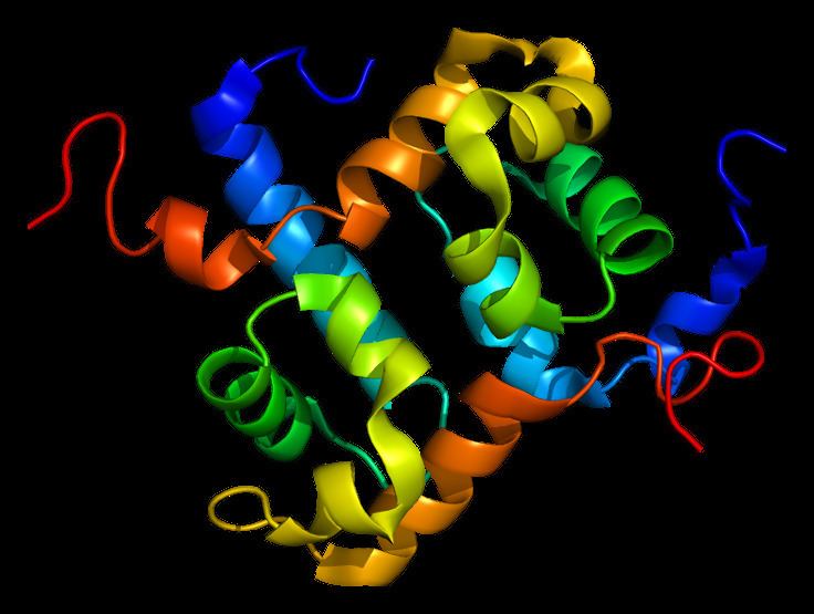
Protein
One ERCC1 molecule and one XPF molecule bind together, forming an ERCC1-XPF heterodimer which is the active nuclease form of the enzyme. In the ERCC1–XPF heterodimer, ERCC1 mediates DNA– and protein–protein interactions. XPF provides the endonuclease active site and is involved in DNA binding and additional protein–protein interactions.
The ERCC4/XPF protein consists of two conserved domains separated by a less conserved region in the middle. The N-terminal region has homology to several conserved domains of DNA helicases belonging to superfamily II, although XPF is not a DNA helicase. The C-terminal region of XPF includes the active site residues for nuclease activity. Most of the ERCC1 protein is related at the sequence level to the C-terminus of the XPF protein, but residues in the nuclease domain are not present. A DNA binding “helix-hairpin-helix” domain at the C-terminus of each protein.
By primary sequence and protein structural similarity, the ERCC1-XPF nuclease is a member of a broader family of structure specific DNA nucleases comprising two subunits. Such nucleases include, for example, the MUS81-EME1 nuclease.
Structure-specific nuclease
The ERCC1–XPF complex is a structure-specific endonuclease. ERCC1-XPF does not cut DNA that is exclusively single-stranded or double-stranded, but it cleaves the DNA phosphodiester backbone specifically at junctions between double-stranded and single-stranded DNA. It introduces a cut in double-stranded DNA on the 5′ side of such a junction, about two nucleotides away. This structure-specificity was initially demonstrated for RAD10-RAD1, the yeast orthologs of ERCC1 and XPF.
The hydrophobic helix–hairpin–helix motifs in the C-terminal regions of ERCC1 and XPF interact to promote dimerization of the two proteins. There is no catalytic activity in the absence of dimerization. Indeed, although the catalytic domain is within XPF and ERCC1 is catalytically inactive, ERCC1 is indispensable for activity of the complex.
Several models have been proposed for binding of ERCC1–XPF to DNA, based on partial structures of relevant protein fragments at atomic resolution. DNA binding mediated by the helix-hairpin-helix domains of ERCC1 and XPF domains positions the heterodimer at the junction between double-stranded and single-stranded DNA.
Nucleotide excision repair
During nucleotide excision repair, several protein complexes cooperate to recognize damaged DNA and locally separate the DNA helix for a short distance on either side of the site of a site of DNA damage. The ERCC1–XPF nuclease incises the damaged DNA strand on the 5′ side of the lesion. During NER, the ERCC1 protein interacts with the XPA protein to coordinate DNA and protein binding.
DNA double-strand break repair
Mammalian cells with mutant ERCC1–XPF are moderately more sensitive than normal cells to agents (such as ionizing radiation) that cause double-stranded breaks in DNA. Particular pathways of both homologous recombination repair and non-homologous end-joining rely on ERCC1-XPF function. The relevant activity of ERCC1–XPF for both types of double-strand break repair is the ability to remove non-homologous 3′ single-stranded tails from DNA ends before rejoining. This activity is needed during a single-strand annealing subpathway of homologous recombination. Trimming of 3’ single-stranded tail is also needed in a mechanistically distinct subpathway of non-homologous end-joining, dependent on the Ku proteins. Homologous integration of DNA, an important technique for genetic manipulation, is dependent on the function of ERCC1-XPF in the host cell.
DNA interstrand crosslink repair
Mammalian cells carrying mutations in ERCC1 or XPF are especially sensitive to agents that cause DNA interstrand crosslinks. Interstrand crosslinks block the progression of DNA replication, and structures at blocked DNA replication forks provide substrates for cleavage by ERCC1-XPF. Incisions may be made on either side of the crosslink on one DNA strand to unhook the crosslink and initiate repair. Alternatively, a double-strand break may be made in the DNA near the ICL, and subsequent homologous recombination repair may involve ERCC1-XPF action. Although not the only nuclease involved, ERCC1–XPF is required for ICL repair during several phases of the cell cycle.
Cerebro-oculo-facio-skeletal syndrome
A few patients with severely disabling ERCC1 mutations that cause cerebro-oculo-facio-skeletal syndrome (COFS) have been reported. COFS syndrome is a rare recessive disorder in which affected individuals undergo rapid neurologic decline and indications of accelerated aging. A very severe case of such disabling mutations is F231L mutation in the tandem helix-hairpin-helix domain of ERCC1 at its interface with XPF. It is shown that this single mutation is very important for the stability of the ERCC1-XPF complex. This Phenylalanine residue is assisting ERCC1 to accommodate a key Phenylalanine residue from XPF (F894) and the mutation (F231L) disturbs this accommodating function. As a consequence, F894 protrudes out of the interface and the mutant complex is dissociating faster compared to the native one. The life span of patients with such mutations is often around 1–2 years.
Cockayne syndrome
One Cockayne syndrome (CS) type II patient designated CS20LO exhibited a homozygous mutation in exon 7 of ERCC1, producing a F231L mutation.
Relevance in chemotherapy
Measuring ERCC1 activity may have utility in clinical cancer medicine because one mechanism of resistance to platinum chemotherapy drugs correlates with high ERCC1 activity. Nucleotide excision repair (NER) is the primary DNA repair mechanism that removes the therapeutic platinum-DNA adducts from the tumor DNA. ERCC1 activity levels, being an important part of the NER common final pathway, may serve as a marker of general NER throughput. This has been suggested for patients with gastric, ovarian, colorectal and bladder cancers. In Non-small cell lung carcinoma (NSCLC), surgically removed tumors that receive no further therapy have a better survival if ERCC1-positive than if ERCC1-negative. Thus ERCC1 positivity is a favorable prognostic marker, referring to how the disease will proceed if not further treated. ERCC1-positive NSCLC tumors do not benefit from adjuvant platinum chemotherapy. However, ERCC1-negative NSCLC tumors, prognostically worse without treatment, derive substantial benefit from adjuvant cisplatin-based chemotherapy. High ERCC1 is thus a negative predictive marker, referring to how it will respond to a specific type of treatment.
ERCC1 genotyping in humans has shown significant polymorphism at codon 118. These polymorphisms may have differential effects on platinum and mitomycin damage.
Deficiency in cancer
ERCC1 protein expression is reduced or absent in 84% to 100% of colorectal cancers, and the promoter of ERCC1 is methylated in 38% of gliomas, resulting in reduced mRNA and protein expression. The promoter of ERCC1 was located in the DNA 5 kilobases upstream of the protein coding region. Frequencies of epigenetic reductions of nine other DNA repair genes have been evaluated in various cancers and range from 2% (OGG1 in papillary thyroid cancer) to 88% and 90% (MGMT in gastric and colon cancers, respectively). Thus, reduction of protein expression of ERCC1 in 84% to 100% of colon cancers indicates that reduced ERCC1 is one of the most frequent reductions of a DNA repair gene observed in a cancer. Deficiency in ERCC1 protein expression appears to be an early event in colon carcinogenesis, since ERCC1 was found to be deficient in 40% of the crypts within 10 cm on each side of colonic adenocarcinomas (within the early field defects from which the cancers likely arose).
Cadmium (Cd) and its compounds are well-known human carcinogens. During Cd-induced malignant transformation, the promoter regions of ERCC1, as well as of hMSH2, XRCC1, and hOGG1, were heavily methylated and both the messenger RNA and proteins of these DNA repair genes were progressively reduced. DNA damage also increased with Cd-induced transformation. Reduction of protein expression of ERCC1 in progression to sporadic cancer is unlikely to be due to mutation. While germ line (familial) mutations in DNA repair genes cause a high risk of cancer (see inherited impairment in DNA repair increases cancer risk), somatic mutations in DNA repair genes, including ERCC1, only occur at low levels in sporadic (non-familial) cancers.
Control of ERCC1 protein level occurred at the translational level. In addition to the wild-type sequence, three splice variants of mRNA ERCC1 exist. ERCC1 mRNA is also found to have either wild-type or three alternative transcription start points. Neither the level of overall mRNA transcription, splice variation nor transcription start point of mRNA correlates with protein level of ERCC1. The rate of ERCC1 protein turnover also does not correlate with ERCC1 protein level. A translational level control of ERCC1, due to a microRNA (miRNA), has been shown during HIV viral infection. A trans-activation response element (TAR) miRNA, coded for by the HIV virus, down-regulates ERCC1 protein expression. TAR miRNA allows ERCC1 mRNA to be transcribed, but acts at the p-body level to prevent translation of ERCC1 protein. (A p-body is a cytoplasmic granule “processing body” that interacts with miRNAs to repress translation or trigger degradation of target RNAs.) In breast cancer cell lines, almost one third (55/167) of miRNA promoters were targets for aberrant methylation (epigenetic repression). In breast cancers themselves, methylation of let-7a-3/let-7b miRNA in particular was found. This indicates that let-7a-3/let-7b can be epigenetically repressed.
Repression of let-7a can cause repression of ERCC1 expression through an intermediary step involving the HMGA2 gene. The let-7a miRNA normally represses the HMGA2 gene, and in normal adult tissues, almost no HMGA2 protein is present. (See also Let-7 microRNA precursor.) Reduction or absence of let-7a miRNA allows high expression of the HMGA2 protein. HMGA proteins are characterized by three DNA-binding domains, called AT-hooks, and an acidic carboxy-terminal tail. HMGA proteins are chromatin architectural transcription factors that both positively and negatively regulate the transcription of a variety of genes. They do not display direct transcriptional activation capacity, but regulate gene expression by changing local DNA conformation. Regulation is achieved by binding to AT-rich regions in the DNA and/or direct interaction with several transcription factors. HMGA2 targets and modifies the chromatin architecture at the ERCC1 gene, reducing its expression. Hypermethylation of the promoter for let-7a miRNA reduces its expression and this allows hyperexpression of HMGA2. Hyperexpression of HMGA2 can then reduce expression of ERCC1.
Thus, there are three mechanisms that may be responsible for the low level of protein expression of ERCC1 in 84% to 100% of sporadic colon cancers. From results in gliomas and in cadmium carcinogenesis, methylation of the ERCC1 promoter may be a factor. One or more miRNAs that repress ERCC1 may be a factor. And epigenetically reduced let-7a miRNA allowing hyperexpression of HMGA2 could also reduce protein expression of ERCC1 in colon cancers. Which epigenetic mechanism occurs most frequently, or whether multiple epigenetic mechanisms reduce ERCC1 protein expression in colon cancers has not been determined.