
 | ||
Dental composite resins are types of synthetic resins which are used in dentistry as restorative material or adhesives. Synthetic resins evolved as restorative materials since they were insoluble, aesthetic, insensitive to dehydration, easy to manipulate and reasonably inexpensive. Composite resins are most commonly composed of Bis-GMA and other dimethacrylate monomers (TEGMA, UDMA, HDDMA), a filler material such as silica and in most current applications, a photoinitiator. Dimethylglyoxime is also commonly added to achieve certain physical properties such as flow ability. Further tailoring of physical properties is achieved by formulating unique concentrations of each constituent.
Contents
- History of use
- Method and clinical application
- Composition
- Advantages
- Disadvantages
- Direct dental composites
- Indirect dental composites
- Direct composite vs amalgam
- Direct vs indirect composites
- References
Many studies have compared the longevity of composite restorations to the longevity of silver-mercury amalgam restorations. Depending on the skill of the dentist, patient characteristics and the type and location of damage, composite restorations can have similar longevity to amalgam restorations. (See Longevity and clinical performance.) In comparison to amalgam, the aesthetics of composite restorations are far superior.
History of use
Traditionally composites set by a chemical setting reaction through polymerization between two pastes. One paste containing an activator (tertiary amine) and the other containing an initiator (benzoyl peroxide). To overcome the disadvantages of this method, such as a short working time, light-curing resin composites were introduced in the 1970’s. The first light-curing units used ultra-violet light to set the material, however this method had a limited curing depth and was a high risk to patients and clinicians. Therefore, UV light-curing units were later replaced by visible light-curing systems which used Camphorquinone as a light source and overcame the issues produced by the UV light-curing units.
The Traditional Period
In the late sixties, composite resins were introduced as an alternative to silicates and unfulfilled resins, which were frequently used by clinicians at the time. Composite resins displayed superior qualities, in that they had higher mechanical properties than silicates and unfulfilled resins. Composite resins were also seen to be beneficial in that the resin would be presented in paste form and, with convenient pressure or bulk insertion technique, would facilitate clinical handling. The faults with composite resins at this time were that they had poor aesthetics, poor marginal adaptation, difficulties with polishing, difficulty with adhesion to the tooth surface, and occasionally, loss of anatomical form.
The Microfilled Period
In 1978, various microfilled systems were introduced into the European market. These composite resins were appealing, in that they were capable of having an extremely smooth surface when finished. These microfilled composite resins also showed a better clinical colour stability and higher resistance to wear than conventional composites, which favoured their aesthetic appearance as well as clinical effectiveness. However, further research showed a progressive weakness in the material over time, leading to micro-cracks and step-like material loss around the composite margin. In 1981, microfilled composites were improved remarkably with regard to marginal retention and adaptation. It was decided, after further research, that this type of composite could be used for most restorations provided the acid etch technique was used and a bonding agent was applied.
The Hybrid Period
Hybrid composites were introduced in the 1980’s and are more commonly known as resin-modified glass ionomer cements (RMGICs). The material consists of a powder containing a radiopaque fluoroaluminosilicate glass and a photoactive liquid contained in a dark bottle or capsule. The material was introduced, as resin composites on their own were not suitable for Class II cavities. RMGICs can be used instead. This mixture or resin and glass ionomer allows the material to be set by light activation (resin), allowing a longer working time. It also has the benefit of the glass ionomer component releasing fluoride and has superior adhesive properties. RMGICs are now recommended over traditional GICs for basing cavities. There is a great difference between the early and new hybrid composites.
Initially, composite restorations in dentistry were very prone to leakage and breakage due to weak compressive strength. In the 1990s and 2000s, composites were greatly improved and have a compression strength sufficient for use in posterior teeth.
Method and clinical application
Today's composite resins have low polymerization shrinkage and low coefficients of thermal shrinkage, which allows them to be placed in bulk while maintaining good adaptation to cavity walls. The placement of composite requires meticulous attention to procedure or it may fail prematurely. The tooth must be kept perfectly dry during placement or the resin will likely fail to adhere to the tooth. Composites are placed while still in a soft, dough-like state, but when exposed to light of a certain blue wavelength (typically 470 nm), they polymerize and harden into the solid filling (for more information, see Light activated resin). It is challenging to harden all of the composite, since the light often does not penetrate more than 2–3 mm into the composite. If too thick an amount of composite is placed in the tooth, the composite will remain partially soft, and this soft unpolymerized composite could ultimately lead to leaching of free monomers with potential toxicity and/or leakage of the bonded joint leading to recurring dental pathology. The dentist should place composite in a deep filling in numerous increments, curing each 2–3 mm section fully before adding the next. In addition, the clinician must be careful to adjust the bite of the composite filling, which can be tricky to do. If the filling is too high, even by a subtle amount, that could lead to chewing sensitivity on the tooth. A properly placed composite is comfortable, aesthetically pleasing, strong and durable, and could last 10 years or more. (By most North American insurance companies 2 years minimum)
The most desirable finish surface for a composite resin can be provided by aluminum oxide disks. Classically, Class III composite preparations were required to have retention points placed entirely in dentin. A syringe was used for placing composite resin because the possibility of trapping air in a restoration was minimized. Modern techniques vary, but conventional wisdom states that because there have been great increases in bonding strength due to the use of dentin primers in the late 1990s, physical retention is not needed except for the most extreme of cases. Primers allow the dentin's collagen fibers to be "sandwiched" into the resin, resulting in a superior physical and chemical bond of the filling to the tooth. Indeed, composite usage was highly controversial in the dental field until primer technology was standardized in the mid to late 1990s. The enamel margin of a composite resin preparation should be beveled in order to improve aesthetics and expose the ends of the enamel rods for acid attack. The correct technique of enamel etching prior to placement of a composite resin restoration includes etching with 30%-50% phosphoric acid and rinsing thoroughly with water and drying with air only. In preparing a cavity for restoration with composite resin combined with an acid etch technique, all enamel cavosurface angles should be obtuse angles. Contraindications for composite include varnish and zinc oxide-eugenol. Composite resins for Class II restorations were not indicated because of excessive occlusal wear in the 1980s and early 1990s. Modern bonding techniques and the increasing unpopularity of amalgam filling material have made composites more attractive for Class II restorations. Opinions vary, but composite is regarded as having adequate longevity and wear characteristics to be used for permanent Class II restorations. Whether composite materials last as long or has the leakage and sensitivity properties when compared to Class II amalgam restorations was described as a matter of debate in 2008.
Composition
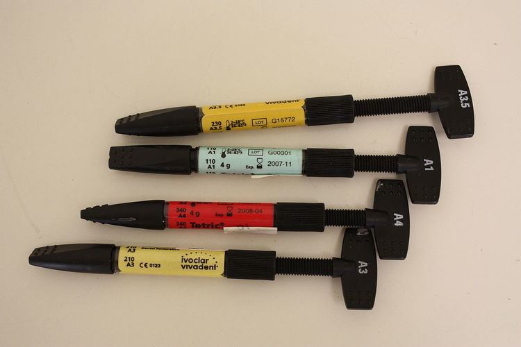
As with other composite materials, a dental composite typically consists of a resin-based oligomer matrix, such as a bisphenol A-glycidyl methacrylate (BISGMA), urethane dimethacrylate (UDMA) or [(semi-crystalline polyceram)] (PEX), and an inorganic filler such as silicon dioxide (silica). Compositions vary widely, with proprietary mixes of resins forming the matrix, as well as engineered filler glasses and glass ceramics. The filler gives the composite wear resistance and translucency. A coupling agent such as silane is used to enhance the bond between these two components. An initiator package (such as: camphorquinone (CQ), phenylpropanedione (PPD) or lucirin (TPO)) begins the polymerization reaction of the resins when external energy (light/heat, etc.) is applied. A catalyst package can control its speed.
Advantages
Advantages of composites:
Disadvantages
Direct dental composites
Direct dental composites are placed by the dentist in a clinical setting. Polymerization is accomplished typically with a hand held curing light that emits specific wavelengths keyed to the initiator and catalyst packages involved. When using a curing light, the light should be held as close to the resin surface as possible, a shield should be placed between the light tip and the operator's eyes. Curing time should be increased for darker resin shades. Light cured resins provide denser restoration than self-cured resins because no mixing is required that might introduce air bubble porosity.
Direct dental composites can be used for:
Indirect dental composites
Indirect composite is cured outside the mouth, in a processing unit that is capable of delivering higher intensities and levels of energy than handheld lights can. Indirect composites can have higher filler levels, are cured for longer times and curing shrinkage can be handled in a better way. As a result, they are less prone to shrinkage stress and marginal gaps and have higher levels and depths of cure than direct composites. For example, an entire crown can be cured in a single process cycle in an extra-oral curing unit, compared to a millimeter layer of a filling.
As a result, full crowns and even bridges (replacing multiple teeth) can be fabricated with these systems.
Indirect dental composites can be used for:
A stronger, tougher and more durable product is expected in principle. But in the case of inlays, not all clinical long-term-studies detect this advantage in clinical practice (see below).
Direct composite vs amalgam
Clinical survival of composite restorations placed in posterior teeth are in the range of amalgam restorations, with some studies seeing a slightly lower or slightly higher survival time compared to amalgam restorations. Improvements in composite technology and application technique make composites a very good alternative to amalgam, while use in large restorations and in cusp capping situations is still debated.
According to a 2012 review article by Demarco et al. covering 34 relevant clinical studies, "90% of the studies indicated that annual failure rates between 1% and 3% can be achieved with Class I and II posterior [rear tooth] composite restorations depending on the definition of failure, and on several factors such as tooth type and location, operator [dentist], and socioeconomic, demographic, and behavioral elements." This compares to a 3% mean annual failure rate reported in a 2004 review article by Manhart et al. for amalgam restorations in posterior stress-bearing cavities.
The Demarco review found that the main reasons cited for failure of posterior composite restorations are secondary caries (i.e. cavities which develop subsequent to the restoration), fracture, and patient behavior, notably bruxism (grinding/clenching.) Causes of failure for amalgam restorations reported in the Manhart et al.review also include secondary caries, fracture (of the amalgam and/or the tooth), as well as cervical overhang and marginal ditching. The Demarco et al. review of composite restoration studies noted that patient factors affect longevity of restorations: Compared to patients with generally good dental health, patients with poorer dental health (possibly due to poor dental hygiene, diet, genetics, frequency of dental checkups, etc.) experience higher rates of failure of composite restorations due to subsequent decay. Socioeconomic factors also play a role: "People who had always lived in the poorest stratus [sic][stratum?] of the population had more restoration failures than those who lived in the richest layer."
The definition of failure applied in clinical studies may affect the reported statistics. Demarco et al note: "Failed restorations or restorations presenting small defects are routinely treated by replacement by most clinicians. Because of this, for many years, the replacement of defective restorations has been reported as the most common treatment in general dental practice..." Demarco et al observe that when both repaired and replaced restorations were classified as failures in one study, the Annual Failure Rate was 1.9%. However, when repaired restorations were reclassified as successes instead of failures, the AFR decreased to 0.7%. Reclassifying repairable minor defects as successes rather than failures is justifiable: "When a restoration is replaced, a significant amount of sound tooth structure is removed and the preparation [i.e. hole] is enlarged". Applying the narrower definition of failure would improve the reported longevity of composite restorations: Composite restorations can often be easily repaired or extended without drilling out and replacing the entire filling. Resin composites will adhere to the tooth and to undamaged prior composite material. In contrast, amalgam fillings are held in place by the shape of the void being filled rather than by adhesion. This means that it is often necessary to drill out and replace an entire amalgam restoration rather than add to the remaining amalgam.
Direct vs indirect composites
It might be expected that the costlier indirect technique leads to a higher clinical performance, however this is not seen in all studies. A study conducted over the course of 11 years reports similar failure rates of direct composite fillings and indirect composite inlays. Another study concludes that although there is a lower failure rate of composite inlays it would be insignificant and anyway too small to justify the additional effort of the indirect technique. Also in the case of ceramic inlays a significantly higher survival rate compared to composite direct fillings can not be detected.
In general, a clear superiority of tooth colored inlays over composite direct fillings could not be established by current review literature (as of 2013).