
TA A03.6.10.003 | FMA 44055 | |
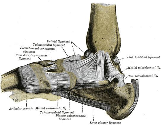 | ||
Latin Ligamentum collaterale mediale articulationis talocruralis, ligamentum deltoideum | ||
The deltoid ligament (or medial ligament of talocrural joint) is a strong, flat, triangular band, attached, above, to the apex and anterior and posterior borders of the medial malleolus. The deltoid ligament is composed of the anterior tibiotalar ligament, tibiocalcaneal ligament, posterior tibiotalar ligament and the tibionavicular ligament. It consists of two sets of fibers, superficial and deep.
Contents
Superficial fibres
Of the superficial fibres,
Deep fibres
The deep fibres (anterior tibiotalar) are attached to the anterior colliculus of the medial malleolus, and below to the anteromedial talus
Coverings
The deltoid ligament is covered by the tendons of the tibialis posterior and flexor digitorum longus.
References
Deltoid ligament Wikipedia(Text) CC BY-SA