
Latin os coccygis TA A02.2.06.001 | MeSH A02.835.232.834.229 FMA 20229 | |
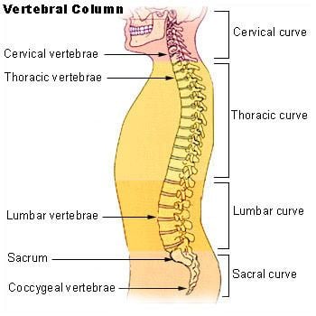 | ||
The coccyx (/ˈkɒksɪks/ KOK-siks; plural: coccyges or coccyxes), commonly referred to as the tailbone, is the final segment of the vertebral column in humans and apes, and certain other mammals such as horses. In animals with bony tails, it is known as tailhead or dock, in bird anatomy as tailfan. It comprises three to five separate or fused coccygeal vertebrae below the sacrum, attached to the sacrum by a fibrocartilaginous joint, the sacrococcygeal symphysis, which permits limited movement between the sacrum and the coccyx.
Contents
Structure
The coccyx is formed of either three, four or five rudimentary vertebrae. It articulates superiorly with the sacrum. In each of the first three segments may be traced a rudimentary body and articular and transverse processes; the last piece (sometimes the third) is a mere nodule of bone. The transverse processes are most prominent and noticeable on the first coccygeal segment. All the segments lack pedicles, laminae and spinous processes. The first is the largest; it resembles the lowest sacral vertebra, and often exists as a separate piece; the remaining ones diminish in size from above downward.
Most anatomy books incorrectly state that the coccyx is normally fused in adults. In fact it has been shown that the coccyx may consist of up to five separate bony segments, the most common configuration being two or three segments.
Surfaces
The anterior surface is slightly concave and marked with three transverse grooves that indicate the junctions of the different segments. It gives attachment to the anterior sacrococcygeal ligament and the levatores ani and supports part of the rectum. The posterior surface is convex, marked by transverse grooves similar to those on the anterior surface, and presents on either side a linear row of tubercles–the rudimentary articular processes of the coccygeal vertebrae. Of these, the superior pair are the largest, and are called the coccygeal cornua they project upward, and articulate with the cornua of the sacrum, and on either side complete the foramen for the transmission of the posterior division of the fifth sacral nerve.
Borders
The lateral borders are thin and exhibit a series of small eminences, which represent the transverse processes of the coccygeal vertebrae. Of these, the first is the largest; it is flattened from before backward, and often ascends to join the lower part of the thin lateral edge of the sacrum, thus completing the foramen for the transmission of the anterior division of the fifth sacral nerve; the others diminish in size from above downward, and are often wanting. The borders of the coccyx are narrow, and give attachment on either side to the sacrotuberous and sacrospinous ligaments, to the coccygeus in front of the ligaments, and to the gluteus maximus behind them.
Apex
The apex is rounded, and has attached to it the tendon of the external anal sphincter. It may be bifid (divided into two).
Extensor coccygis
The extensor coccygis is a slender muscle fascicle, which is not always present. It extends over the lower part of the posterior surface of the sacrum and coccyx. It arises by tendinous fibers from the last segment of the sacrum, or first piece of the coccyx, and passes downward to be inserted into the lower part of the coccyx. It is a rudiment of the extensor muscle of the caudal vertebrae of other animals.
Sacrococcygeal and intercoccygeal joints
The joints are variable and may be: (1) synovial joints; (2) thin discs of fibrocartilage; (3) intermediate between these two; (4) ossified.
Function
In humans and other tailless primates (e.g., great apes) since Nacholapithecus (a Miocene hominoid), the coccyx is the remnant of a vestigial tail, but still not entirely useless; it is an important attachment for various muscles, tendons and ligaments—which makes it necessary for physicians and patients to pay special attention to these attachments when considering surgical removal of the coccyx. Additionally, it is also a part of the weight-bearing tripod structure which acts as a support for a sitting person. When a person sits leaning forward, the ischial tuberosities and inferior rami of the ischium take most of the weight, but as the sitting person leans backward, more weight is transferred to the coccyx.
The anterior side of the coccyx serves for the attachment of a group of muscles important for many functions of the pelvic floor (i.e., defecation, continence, etc.): the levator ani muscle, which include coccygeus, iliococcygeus, and pubococcygeus. Through the anococcygeal raphe, the coccyx supports the position of the anus. Attached to the posterior side is gluteus maximus which extend the thigh during ambulation.
Many important ligaments attach to the coccyx: the anterior and posterior sacrococcygeal ligaments are the continuations of the anterior and posterior longitudinal ligaments that stretches along the entire spine. Additionally, the lateral sacrococcygeal ligaments complete the foramina for the last sacral nerve. And, lastly, some fibers of the sacrospinous and sacrotuberous ligaments (arising from the spine of the ischium and the ischial tuberosity respectively) also attach to the coccyx.
An extension of the pia mater, the filum terminale, extends from the apex of the conus, and inserts on the coccyx.
Clinical significance
Injuring the coccyx can give rise to a painful condition called coccydynia and one or more of the bones or the connections thereof may be broken, fractured tailbone. A number of tumors are known to involve the coccyx; of these, the most common is sacrococcygeal teratoma. Both coccydynia and coccygeal tumors may require surgical removal of the coccyx (coccygectomy). One very rare complication of coccygectomy is a type of perineal hernia known as a coccygeal hernia.
Etymology
The term coccyx is derived from the ancient Greek word κόκκυξ kokkyx "cuckoo"; the latter is attested in the writings of the Greek physician Herophilus to denote the end of the vertebral column. This Greek name for the cuckoo was applied as the last three or four bones of the coccyx resemble the beak of this bird, when viewed from the side.
This established etymological explanation can also be found in the writings of the 16th century anatomist Andreas Vesalius whom wrote: os cuculi, a similitudine rostri cuculi avis (the cuckoo bone shows a likeness to the beak of the cuckoo bird). Vesalius used the Latin expression os cuculi, with os, bone and cuculus, the Latin name for the cuckoo. The 16th/17th century French anatomist Jean Riolan the Younger gives a rather hilarious etymological explanation, as he writes: quia crepitus, qui per sedimentum exeunt, ad is os allisi, cuculi vocis similitudinem effingunt (because the sound of the farts that leave the anus and dash against this bone, shows a likeness to the call of the cuckoo). The latter is not considered as potential candidate.
Besides os cuculi, os caudae, with caudae, of the tail is attested. This Latin expression might be the source of the English, French language, German and Dutch terms tailbone, l'os de la queue, Schwanzbein and staartbeen. In the current official anatomic Latin nomenclature, Terminologia Anatomica, coccyx and os coccygis is used.