
Entrez 3381 | Ensembl ENSG00000029559 | |
 | ||
External IDs OMIM: 147563 MGI: 96389 HomoloGene: 3644 GeneCards: IBSP | ||
Bone sialoprotein (BSP) is a component of mineralized tissues such as bone, dentin, cementum and calcified cartilage. BSP is a significant component of the bone extracellular matrix and has been suggested to constitute approximately 8% of all non-collagenous proteins found in bone and cementum. BSP, a SIBLING protein, was originally isolated from bovine cortical bone as a 23-kDa glycopeptide with high sialic acid content.
Contents
The human variant of BSP is called bone sialoprotein 2 also known as cell-binding sialoprotein or integrin-binding sialoprotein and is encoded by the IBSP gene.
Structure
Native BSP has an apparent molecular weight of 60-80 kDa based on SDS-PAGE, which is a considerable deviation from the predicted weight (based on cDNA sequence) of approximately 33 kDa. The mammalian BSP cDNAs encode for proteins averaging 327 amino acids, which includes the 16-residue preprotein secretory signal peptide. Among the mammalian cDNAs currently characterized, there is an approximate 45% conservation of sequence identity and a further 10-23% conservative substitution. The protein is highly acidic (pKa of ~ 3.9) and contains a large amount of Glu residues, constituting ~22% of the total amino acid.
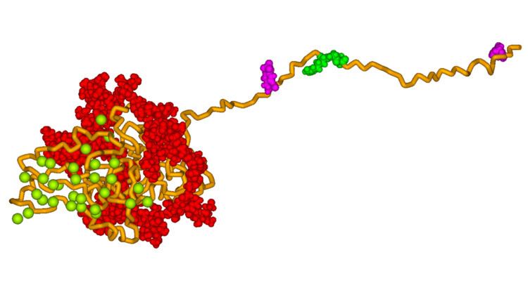
Secondary structure prediction and hydrophobicity analyses suggest that the primary sequence of BSP has an open, flexible structure with the potential to form regions of α-helix and some β-sheet. However, the majority of studies have demonstrated that BSP has no α-helical or β-sheet structure by 1D NMR and circular dichroism. Analysis of native protein by electron microscopy confirm that the protein has an extended structure approximately 40 nm in length. This flexible conformation suggests that the protein has few structural domains, however it has been suggested that there may be several spatially segmented functional domains including a hydrophobic collagen-binding domain (rattus norvegicus residues 36-57), a hydroxyapatite-nucleating region of contiguous glutamic acid residues (rattus norvegicus residues 78-85, 155-164) and a classical integrin-binding motif (RGD) near the C-terminal (rattus norvegicus residues 288-291).
BSP has been demonstrated to be extensively post-translationally modified, with carbohydrates and other modifications comprising approximately 50% of the molecular weight of the native protein. These modifications, which include N- and O-linked glycosylation, tyrosine sulfation and serine and threonine phosphorylation, make the protein highly heterogeneous.
Function
The amount of BSP in bone and dentin is roughly equal, however the function of BSP in these mineralized tissues is not known. One possibility is that BSP acts as a nucleus for the formation of the first apatite crystals. As the apatite forms along the collagen fibres within the extracellular matrix, BSP could then help direct, redirect or inhibit the crystal growth.
Additional roles of BSP are angiogenesis and protection from complement-mediated cell lysis. Regulation of the BSP gene is important to bone matrix mineralization and tumor growth in bone.