
Latin caementum | FMA 55630 | |
 | ||
TA A05.1.03.057A03.1.03.007 | ||
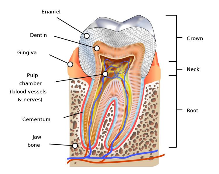
Cementum is a specialized calcified substance covering the root of a tooth. The cementum is the part of the periodontium that attaches the teeth to the alveolar bone by anchoring the periodontal ligament.
Contents
Development
Cementum is excreted by cells called cementoblasts within the root of the tooth and is thickest at the root apex. These cementoblasts develop from undifferentiated mesenchymal cells in the connective tissue of the dental follicle or sac.
Unlike ameloblasts and odontoblasts, which leave no cellular bodies in their secreted products, during the later steps within the stage of apposition, many of the cementoblasts become entrapped by the cementum they produce, becoming cementocytes. Thus again, cementum is more similar to alveolar bone, with its osteoblasts becoming entrapped osteocytes.
Cementum is capable of repairing itself to a limited degree and is not resorbed under normal conditions.
Features
Cementum is slightly softer than dentin and consists of about 45% to 50% inorganic material (hydroxylapatite) by weight and 50% to 55% organic matter and water by weight. The organic portion is composed primarily of collagen and proteoglycans. Cementum is avascular, receiving its nutrition through its own imbedded cells from the surrounding vascular periodontal ligament.
The cementum is light yellow and slightly lighter in color than dentin. It has the highest fluoride content of all mineralized tissue. Cementum also is permeable to a variety of materials. It is formed continuously throughout life because a new layer of cementum is deposited to keep the attachment intact as the superficial layer of cementum ages. Cementum on the root ends surrounds the apical foramen and may extend slightly onto the inner wall of the pulp canal.
Structure
The cells of cementum are the entrapped cementoblasts, the cementocytes. Each cementocyte lies in its lacuna, similar to the pattern noted in bone. These lacunae also have canaliculi or canals. Unlike those in bone, however, these canals in cementum do not contain nerves, nor do they radiate outward. Instead, the canals are oriented toward the periodontal ligament and contain cementocytic processes that exist to diffuse nutrients from the ligament because it is vascularized.
After the apposition of cementum in layers, the cementoblasts that do not become entrapped in cementum line up along the cemental surface along the length of the outer covering of the periodontal ligament. These cementoblasts can form subsequent layers of cementum if the tooth is injured.
Sharpey fibers are part of the principal collagenous fibers of the periodontal ligament embedded in the cementum and alveolar bone to attach the tooth to the alveolus.
Cementoenamel junction
The cementum joins the enamel to form the cementoenamel junction (CEJ), which is referred to as the cervical line.
Three possible types of transitional interfaces may be present at the CEJ. The traditional view was that certain interfaces dominated in certain oral cavities. The CEJ may exhibit all of these interfaces in an individual’s oral cavity, and there is even considerable variation when one tooth is traced circumferentially.
Dentinocemental junction
When the cementoid reaches the full thickness needed, the cementoid surrounding the cementocytes becomes mineralized, or matured, and is then considered cementum. Because of the apposition of cementum over the dentin, the dentinocemental junction (DCJ) is formed. This interface is not as defined, either clinically or histologically, as that of the dentinoenamel junction (DEJ), given that cementum and dentin are of common embryological background, unlike that of enamel and dentin.
The dentinocemental junction (DCJ) is a relatively smooth area in the permanent tooth, and attachment of cementum to the dentin is firm but not understood completely.
Types
Two kinds of cementum are formed: acellular and cellular, and fibers can be intrinsic or extrinsic, resulting in four possible permutations; the first cementum to be formed during tooth development is acellular extrinsic fiber cementum. The acellular layer of cementum is living tissue that does not incorporate cells into its structure and usually predominates on the coronal half of the root; cellular cementum occurs more frequently on the apical half.
Pathology
DNA studies
A 2010 archeological study has found that cementum has five times the amount of mitochondrial DNA compared to dentin, which is commonly sampled. Teeth are increasingly utilized as a source of nuclear DNA to aid identification of human remains. DNA extraction and the results of genetic analysis from the tissue are extremely variable and to some extent unpredictable. However, the quantity of DNA available in dentin is affected by age and dental disease, whereas that in cementum is not.