
ICD-9-CM 92.14 MedlinePlus 003833 | OPS-301 code 3-705 | |
 | ||
A bone scan or bone scintigraphy /sɪnˈtɪɡrəfi/ is a nuclear medicine imaging technique of the bone. It can help diagnose a number of bone conditions, including; cancer of the bone or metastasis, location of bone inflammation and fractures (that may not be visible in traditional X-ray images), and bone infection.
Contents
Nuclear medicine provides functional imaging and allows visualisation of bone metabolism or bone remodeling, which most other imaging techniques (such as X-ray computed tomography, CT) cannot. Bone scintigraphy competes with positron emission tomography (PET) for imaging of abnormal metabolism in bones, but is considerably less expensive. Bone scintigraphy has higher sensitivity but lower specificity than CT and MRI for diagnosis of scaphoid fractures following negative plain radiography.
History
Some of the earliest investigations into skeletal metabolism were carried out by George de Hevesy in the 1930s, using Phosphorus-32. In the 1950s and 1960s Calcium-45 was investigated, but as a beta emitter proved difficult to image. Imaging of positron and gamma emitters such as Fluorine-18 and isotopes of strontium with rectilinear scanners was more useful. Use of Technetium-99m (99mTc) labelled phosphates, diphophonates or similar agents, as in the modern technique, was first proposed in 1971.
Principle
The most common radiopharmaceutical for bone scintigraphy is 99mTc with methylene diphosphonate (MDP). MDP adsorbs onto the crystalline hydroxyapatite mineral of bone. Mineralisation occurs at osteoblasts, representing sites of bone growth, where MDP (and other diphosphates) "bind to the hydroxyapatite crystals in proportion to local blood flow and osteoblastic activity and are therefore markers of bone turnover and bone perfusion".
The more active the bone turnover, the more radioactive material will be seen. Some tumors, fractures and infections show up as areas of increased uptake.
Technique
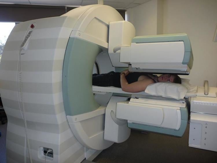
In a typical bone scan technique, the patient is injected (usually into a vein in the arm or hand, occasionally the foot) with up to 740 MBq of technetium-99m-MDP and then scanned with a gamma camera, which captures planar anterior and posterior or single photon emission computed tomography (SPECT) images. In order to view small lesions SPECT imaging technique may be preferred over planar scintigraphy.
In a single phase protocol (skeletal imaging alone), which will primarily highlight osteoblasts, images are usually acquired 2-5 hours after the injection (after four hours 50-60% of the activity will be fixed to bones). A two or three phase protocol utilises additional scans at different points after the injection to obtain additional diagnostic information. A dynamic (i.e. multiple acquired frames) study immediately after the injection captures perfusion information. A second phase "blood pool" image following the perfusion (if carried out in a three phase technique)can help to diagnose inflammatory conditions or problems of blood supply.
A typical effective dose obtained during a bone scan is 6.3 millisieverts (mSv).