
 | ||
Similar Epiphysis, Femur, Tibia | ||
Bone age assessment
Bone age is the degree of maturation of a child's bones. As a person grows from fetal life through childhood, puberty, and finishes growth as a young adult, the bones of the skeleton change in size and shape. These changes can be seen by x-ray. The "bone age" of a child is the average age at which children reach this stage of bone maturation. A child's current height and bone age can be used to predict adult height. For most people, their bone age is the same as their biological age but for some individuals, their bone age is a couple years older or younger. Those with advanced bone ages typically hit a growth spurt early on but stop growing early sooner while those with delayed bone ages hit their growth spurt later than normal. Kids who are below average height do not necessarily have a delayed bone age; in fact their bone age could actually be advanced which if left untreated, will stunt their growth.
Contents
- Bone age assessment
- Bone age made easy
- Methods
- Height prediction
- Clinical application of bone age readings
- References
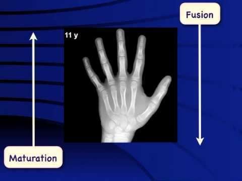
At birth, only the metaphyses of the "long bones" are present. The long bones are those that grow primarily by elongation at an epiphysis at one end of the growing bone. The long bones include the femurs, tibias, and fibulas of the lower limb, the humeri, radii, and ulnas of the upper limb (arm + forearm), and the phalanges of the fingers and toes. The long bones of the leg comprise nearly half of adult height. The other primary skeletal component of height is the spine and skull.
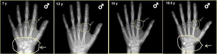
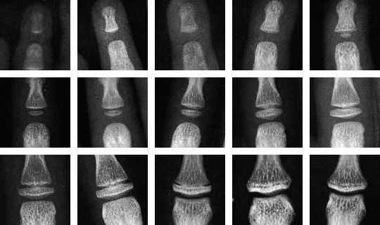
As a child grows the epiphyses become calcified and appear on the x-rays, as do the carpal and tarsal bones of the hands and feet, separated on the x-rays by a layer of invisible cartilage where most of the growth is occurring. As sex steroid levels rise during puberty, bone maturation accelerates. As growth nears conclusion and attainment of adult height, bones begin to approach the size and shape of adult bones. The remaining cartilaginous portions of the epiphyses become thinner. As these cartilaginous zones become obliterated, the epiphyses are said to be "closed" and no further lengthening of the bones will occur. A small amount of spinal growth concludes an adolescent's growth.
Pediatric endocrinologists frequently order bone age x-rays to evaluate children for advanced or delayed growth and physical development. These are interpreted by pediatric radiologists, physicians who are experts in using medical imaging for pediatric diagnosis and therapy.
Bone age made easy
Methods
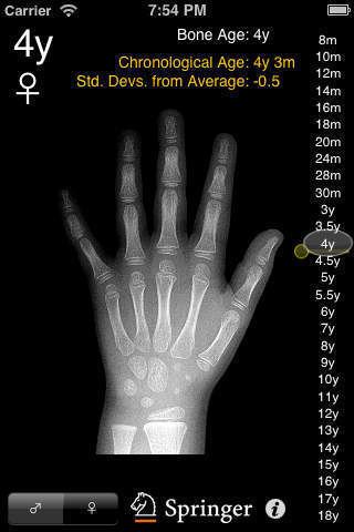
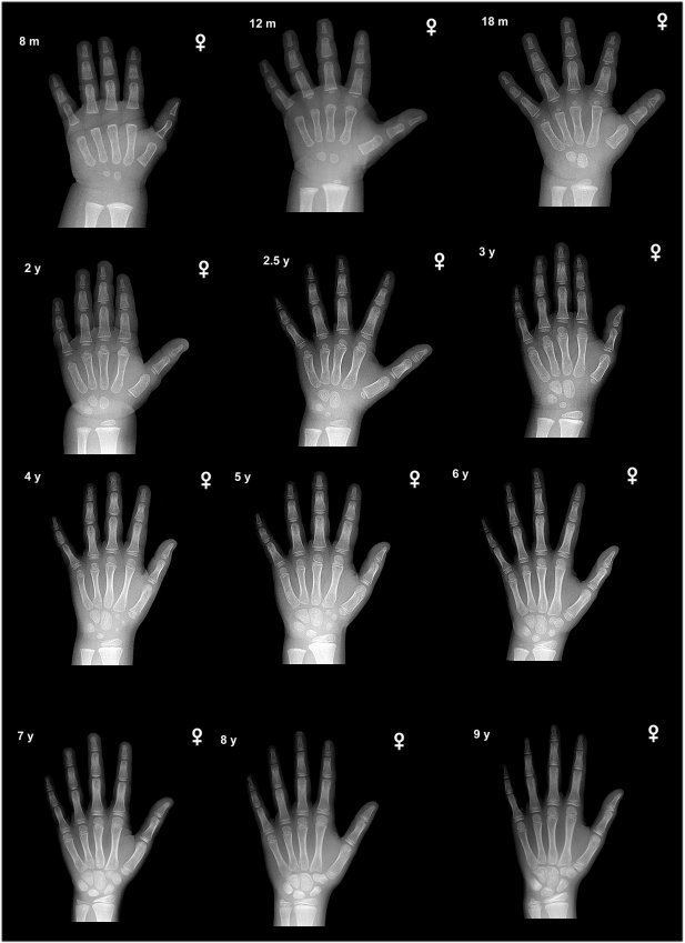
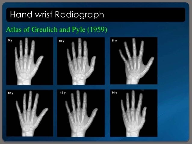
The most commonly used method is based on a single x-ray of the left hand, fingers, and wrist. A hand is easily x-rayed with minimal radiation and shows many bones in a single view. The bones in the x-ray are compared to the bones of a standard atlas, usually "Greulich and Pyle".
A more complex method also based on hand x-rays is the "TW2" or the "TW3" method (TW = Tanner Whitehouse) method.
An atlas based on knee maturation has also been compiled.
Bone age rating is a procedure well-suited for automation by computer. The main advantage is the elimination of the variability of rating between different human raters. The BoneXpert method is the most prominent example of a completely automated method.
The hands of infants do not change much in the first year of life and if precise bone age assessment is desired, an x-ray of approximately half of the skeleton (a "hemiskeleton" view) may be obtained to assess some of the areas such as shoulders and pelvis which change more in infancy.
Height prediction
Statistics have been compiled to indicate the percentage of height growth remaining at a given bone age. By simple arithmetic, a predicted adult height can be computed from a child's height and bone age. Separate tables are used for boys and girls because of the sex difference in timing of puberty, and slightly different percentages are used for children with unusually advanced or delayed bone maturation. These tables, the Bayley-Pinneau tables, are included as an appendix in the Greulich and Pyle atlas.
In a number of conditions involving atypical growth, bone age height predictions are less accurate. For example, in children born small for gestational age who remain short after birth, the bone age is a poor predictor of adult height.
Clinical application of bone age readings
An advanced or delayed bone age does not always indicate disease or "pathologic" growth. Conversely, the bone age may be normal in some conditions of abnormal growth. Children do not mature at exactly the same time. Just as there is wide variation among the normal population in age of losing teeth or experiencing the first menstrual period, the bone age of a healthy child may be a year or two advanced or delayed. Those with an advanced bone age typically hit a growth spurt early on but stop growing at an earlier age. Consequently, when a naturally short child has an advanced bone age, it stunts their growth at an early age leaving them even shorter than they would have been. Because of this, those who are short with an advanced bone age, need medical attention before their bones fully fuse.
An advanced bone age is common when a child has had prolonged elevation of sex steroid levels, as in precocious puberty or congenital adrenal hyperplasia.
Bone age tends to be slightly advanced in cases of premature adrenarche, particularly in children who have been overweight from a young age or those with lipodystrophy. Individuals with an advanced bone age usually experience an early growth spurt but cease growing at a younger age than typical.
Bone age may be significantly advanced in genetic overgrowth syndromes, such as Sotos syndrome, Beckwith-Wiedemann syndrome and Marshall-Smith syndrome.
Bone maturation is delayed with the variation of normal development termed Constitutional delay of growth and puberty, but delay also accompanies growth failure due to growth hormone deficiency and hypothyroidism.