
Symbol SAG Entrez 6295 OMIM 181031 | Alt. symbols arrestin-1 HUGO 10521 RefSeq NM_000541 | |
 | ||
Arrestins are a small family of proteins important for regulating signal transduction at G protein-coupled receptors. Arrestins were first discovered as a part of a conserved two-step mechanism for regulating the activity of G protein-coupled receptors (GPCRs) in the visual rhodopsin system by Hermann Kühn and co-workers and in the β-adrenergic system by Martin J. Lohse and co-workers.
Contents
Function
In response to a stimulus, GPCRs activate heterotrimeric G proteins. In order to turn off this response, or adapt to a persistent stimulus, active receptors need to be desensitized. The first step is phosphorylation by a class of serine/threonine kinases called G protein coupled receptor kinases (GRKs). GRK phosphorylation specifically prepares the activated receptor for arrestin binding. Arrestin binding to the receptor blocks further G protein-mediated signaling and targets receptors for internalization, and redirects signaling to alternative G protein-independent pathways, such as β-arrestin signaling. In addition to GPCRs, arrestins bind to other classes of cell surface receptors and a variety of other signaling proteins.
Subtypes
Mammals express four arrestin subtypes and each arrestin subtype is known by multiple aliases. The systematic arrestin name (1-4) plus the most widely used aliases for each arrestin subtype are listed in bold below:
Fish and other vertebrates appear to have only three arrestins: no equivalent of arrestin-2, which is the most abundant non-visual subtype in mammals, was cloned so far. The proto-chordate C. intestinalis (sea squirt) has only one arrestin, which serves as visual in its mobile larva with highly developed eyes, and becomes generic non-visual in the blind sessile adult. Conserved positions of multiple introns in its gene and those of our arrestin subtypes suggest that they all evolved from this ancestral arrestin. Lower invertebrates, such as roundworm C. elegans, also have only one arrestin. Insects have arr1 and arr2, originally termed “visual arrestins” because they are expressed in photoreceptors, and one non-visual subtype (kurtz in Drosophila). Later arr1 and arr2 were found to play an important role in olfactory neurons and renamed “sensory”. Fungi have distant arrestin relatives involved in pH sensing.
Tissue distribution
One or more arrestin is expressed in virtually every eukaryotic cell. In mammals, arrestin-1 and arrestin-4 are largely confined to photoreceptors, whereas arrestin-2 and arrestin-3 are ubiquitous. Neurons have the highest expression level of both non-visual subtypes. In neuronal precursors both are expressed at comparable levels, whereas in mature neurons arrestin-2 is present at 10-20 fold higher levels than arrestin-3.
Mechanism
Arrestins block GPCR coupling to G proteins in two ways. First, arrestin binding to the cytoplasmic face of the receptor occludes the binding site for heterotrimeric G-protein, preventing its activation (desensitization). Second, arrestin links the receptor to elements of the internalization machinery, clathrin and clathrin adaptor AP2, which promotes receptor internalization via coated pits and subsequent transport to internal compartments, called endosomes. Subsequently, the receptor could be either directed to degradation compartments (lysosomes) or recycled back to the plasma membrane where it can again signal. The strength of arrestin-receptor interaction plays a role in this choice: tighter complexes tend to increase the probability of receptor degradation (Class B), whereas more transient complexes favor recycling (Class A), although this “rule” is far from absolute.
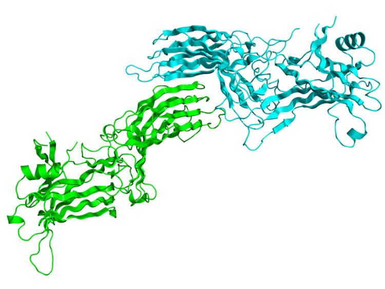
Structure
Arrestins are elongated molecules, in which several intra-molecular interactions hold the relative orientation of the two domains. In unstimulated cell arrestins are localized in the cytoplasm in this basal “inactive” conformation. Active phosphorylated GPCRs recruit arrestin to the plasma membrane. Receptor binding induces a global conformational change that involves the movement of the two arrestin domains and the release of its C-terminal tail that contains clathrin and AP2 binding sites. Increased accessibility of these sites in receptor-bound arrestin targets the arrestin-receptor complex to the coated pit. Arrestins also bind microtubules (part of the cellular “skeleton”), where they assume yet another conformation, different from both free and receptor-bound form. Microtubule-bound arrestins recruit certain proteins to the cytoskeleton, which affects their activity and/or redirects it to microtubule-associated proteins.
Arrestins shuttle between cell nucleus and cytoplasm. Their nuclear functions are not fully understood, but it was shown that all four mammalian arrestin subtypes remove some of their partners, such as protein kinase JNK3 or the ubiquitin ligase Mdm2, from the nucleus. Arrestins also modify gene expression by enhancing transcription of certain genes.