
 | ||
Latin Os tibiale, os tibiale externum or naviculare secundarium | ||
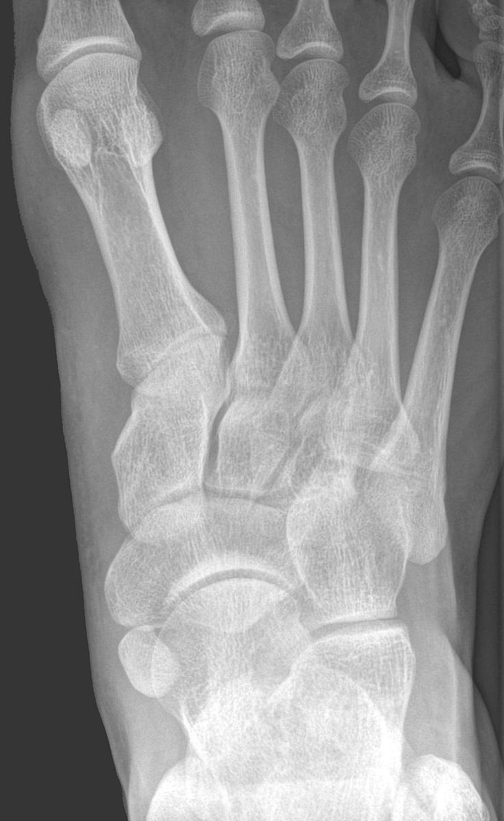
An accessory navicular bone is an accessory bone of the foot that occasionally develops abnormally in front of the ankle towards the inside of the foot. This bone may be present in approximately 2-21% of the general population and is usually asymptomatic. When it is symptomatic, surgery may be necessary.
Contents
Surgery can be performed at any age because it does not alter any other bones.
Symptoms of an accessory navicular bone may include plantar fasciitis, bunions and heel spurs.
Classification
The Geist classification divides the accessory navicular bones into three types.
Treatment
Aside from surgery, there are a few options for handling an accessory navicular bone that has become symptomatic. This includes immobilization, icing, medicating, physical therapy, and orthotic devices. Immobilizing involves placing the foot and ankle in a cast or removable walking boot. This alleviates stressors on the foot and can decrease inflammation. Icing will help reduce swelling and inflammation. Medication involves usage of nonsteroidal anti-inflammatory drugs, or steroids (taken orally or injected) to decrease inflammation. Physical therapy can be prescribed in order to strengthen the muscles and help decrease inflammation. Physical therapy can also help prevent the symptoms from returning. Orthotic devices (arch support devices that fit in a shoe) can help prevent future symptoms. Occasionally, the orthotic device will dig into the edge of the accessory navicular and cause discomfort. For this reason, the orthotic devices made for the patient should be carefully constructed.