
 | ||
Osteoarthritis is a group of mechanical abnormalities resulting in joint destruction, which can occur in the wrist. These abnormalities include degeneration of cartilage and hypertrophic bone changes, which can lead to pain, swelling and loss of function. Osteoarthritis of the wrist is one of the most common conditions seen by hand surgeons.
Contents
- Signs and symptoms
- Cause
- SLAC
- SNAC
- Stages
- Mechanism
- The wrist
- Proximal row
- Distal row
- Diagnosis
- Medical history
- Physical examination
- X rays
- Treatment
- Stage I
- Stage II
- Stage III
- Stage IV
- References
Osteoarthritis of the wrist can be idiopathic, but it is mostly seen as a post-traumatic condition. There are different types of post-traumatic osteoarthritis. Scapholunate Advanced Collapse (SLAC) is the most common form, followed by Scaphoid Non-union Advanced Collapse (SNAC). Other post-traumatic causes such as intra-articular fractures of the distal radius or ulna can also lead to wrist osteoarthritis, but are less common.
Signs and symptoms
The most common initial symptom of wrist osteoarthritis is joint pain. The pain is brought on by activity and increases when there is activity after resting. Other signs and symptoms, as with any joint affected by osteoarthritis, include:
These symptoms can lead to loss of function and less daily activity.
Cause
SLAC and SNAC are both caused by injury, for example a fall on an extended hand. SLAC is caused by rupture of the scapholunate ligament, SNAC is caused by a scaphoid fracture which does not heal and because of that will develop in a non-union fracture. SLAC is more common than SNAC; 55% of the patients with wrist osteoarthritis has a SLAC wrist. Although they have a different underlying pathology, they both lead to abnormal wrist kinematics which will eventually lead to osteoarthritis of the wrist.
SLAC
Rupture of the scapholunate ligament changes wrist joint kinematics. Due to lack of stability from the separated scaphoid and lunate, the lunate may angulate to the posterior side of the hand. This is known as a dorsal intercalated segment instability deformity (DISI), which disrupts the perfect fit between the radius and the scaphoid carpal bone. This process is known as SLAC and eventually will lead to degenerative osteoarthritis of the radioscaphoid joint.
SNAC
Scaphoid non-union fractures changes scaphoid bone shape, which leads to abnormal joint kinematics. Due to lack of stability from the distorted scaphoid, a DISI can be developed. This process is known as SNAC. As in SLAC, this will lead to degenerative osteoarthritis.
Stages
Post-traumatic osteoarthritis can be classified into four stages. These stages are similar between SLAC and SNAC wrists. Each stage has a different treatment.
Mechanism
In order to understand the cause of post-traumatic wrist osteoarthritis it is important to know and understand the anatomy of the wrist. The hand is subdivided into three parts:
The wrist
The wrist consists of eight small carpal bones. Each of these carpal bones has a different size and shape. They contribute towards the stability of the wrist and are ranked in two rows, each consisting of four bones.
Proximal row
From lateral to medial and when viewed from anterior, the proximal row is formed by the:
Distal row
From lateral to medial and when viewed from anterior, the distal row is formed by the:
Diagnosis
Osteoarthritis of the wrist is predominantly a clinical diagnosis, and thus is primarily based on the patients medical history, physical examination and wrist X-rays.
Medical history
Medical history of the patient should include age, hand dominance, occupation and most important an evaluation of recent hand traumas.
Physical examination
Examination will often show tenderness at the radioscaphoid joint (when palpated or while moving the radioscaphoid joint), dorsal radial swelling and instability of the wrist joint. Notice that people may say they have trouble with rising from a chair when pressure is exerted on the hands by pushing against the handrail. Younger people may complain about not being able to do push-ups anymore because of a painful hand.
There are a number of tests and actions that can be performed when a patient is suspected of having osteoarthritis caused by SLAC or SNAC.
SLAC:
Tests:
SNAC:
X-rays
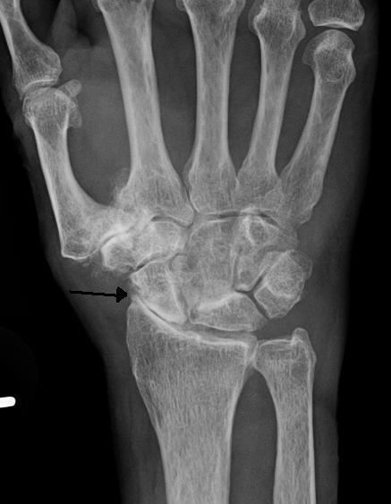
Osteoarthritis between the radius bone and the carpals is indicated by a radiocarpal joint space of less than 2mm.
X-rays can be very helpful in diagnosing and differentiating between SNAC and SLAC wrists. On the other hand, X-rays are not always sufficient to distinguish between different stages. It is important to note that both hands need to be compared. Therefore, two X-rays are needed: one from the left and one from the right hand. When the X-ray is inconclusive, wrist arthroscopy can be performed.
SLAC
Because the scapholunate ligament is ruptured, the scaphoid and lunate are not longer connected. This results in a larger space between the two bones, also known as the Terry Thomas sign. A space larger than 3 mm is suspicious and a space larger than 5 mm is a proven SLAC pathology. Scaphoid instability due to the ligament rupture can be stactic or dynamic. When the X-ray is diagnostic and there is a convincing Terry Thomas sign it is a static scaphoid instability. When the scaphoid is made unstable by either the patient or by manipulation by the examining physician it is a dynamic instability.
In order to diagnose a SLAC wrist you need a posterior anterior (PA) view X-ray, a lateral view X-ray and a fist view X-ray. The fist X-ray is often made if there is no convincing Terry Thomas sign. A fist X-ray of a scapholunate ligament rupture will show a descending capitate. Making a fist will give pressure at the capitate, which will descend if there is a rupture in the scapholunate ligament.
SNAC
In order to diagnose a SNAC wrist you need a PA view X-ray and a lateral view X-ray. As in SLAC, the lateral view X-ray is performed to see if there is a DISI. Computed tomography (CT) or Magnetic Resonance Imaging (MRI) are rarely used to diagnose SNAC or SLAC wrist osteoarthritis because there is no additional value. Also, these techniques are much more expensive than a standard X-ray. CT or MRI may be used if there is a strong suspicion for another underlying pathology or disease.
Treatment
Post-traumatic wrist osteoarthritis can be treated conservatively or with a surgical intervention. In many patients, a conservative (non-surgical) approach is sufficient. Because osteoarthritis is progressive and symptoms may get worse, surgical treatment is advised in any stage.
Stage I
For stage I, normally, nonsurgical treatment is sufficient. This type of therapy includes the use of splint or cast immobilization, injections of corticosteroid in the pain causing joints and the use of a systemic non-steroidal anti-inflammatory drug to reduce pain and improve the functional use of the affected joint. However, the amount of pain that can be suppressed by nonsurgical therapy is limited and with the progression of the wrist osteoarthritis surgical treatment is inevitable.
In stage I surgical treatment often consists of neurectomy of the posterior interosseous nerve and is often combined with other procedures. In the case of a SLAC, the scapholunate ligament can be reconstructed in combination with a radial styloidectomy, in which the radial styloid is surgically removed from the distal radius. In the case of a SNAC, the scaphoid can be reconstructed by fixating the scaphoid with a screw or by placing a bone graft(Matti-Russe procedure)to increase the stability of the scaphoid.
Stage II
In the treatment of stage II wrist osteoarthritis, there are two treatment options that have proved to be most successful. The first treatment option is proximal row carpectomy. During this surgical intervention the proximal row of the carpal bones is removed (scaphoid, lunate, triquetrum, pisiform). It is important that the radioscaphocapitate ligament is left intact, because if the ligament is not preserved the capitate bone will translate to the ulnar side of the wrist and move away from the distal radius. The new formed joint between the capitate and the lunate fossa of the distal radius is not as congruent as the former scaphoid-lunate-radius joint, however the results of proximal row carpectomy are generally excellent. In patients older than 40 years proximal row carpectomy is preferred because these patients have a small chance of developing osteoarthritis in the new formed capitate-radial joint during their remaining life.
Patients younger than 40 years have a big chance to develop osteoarthritis in the radiocapitate joint. These patients have longer to live, therefore the incongruence of the joint will exist for a longer time. Thus, in this patient population four-corner arthrodesis is the treatment of first choice. The capitate, lunate, hamate and triquetrum are bounded together in this procedure and the scaphoid is excised. Before the arthrodesis is executed, the lunate must be reduced out of DISI position. Because the radiolunate joint is typically preserved in stage II SLAC and SNAC wrists, this joint can be the only remaining joint of the proximal wrist. Both procedures are often combined with wrist denervation, as described in the text of treatment stage I.
Stage III
The only treatment option for stage III wrist osteoarthritis is four-corner arthrodesis, as described above in stage II. Proximal row carpectomy is not an option, because in stage III patients the capitate is already affected by the osteoarthritis. So, this procedure would merely lead to a new painful joint.
Stage IV
In this stage there are two surgical treatment options; total wrist arthroplasty and total wrist arthrodesis. Total wrist arthrodesis has become the standard surgical treatment for patients with stage IV wrist osteoarthritis. During this procedure the carpal bones are all fused together and are then fastened to the distal radius. Patients who still want to undertake heavy labor benefit the most of this surgical approach, because after surgery and recovery this is still possible. However, the arc of motion is extremely diminished by this type of surgery.
The best option for those who wish for a motion-sparing procedure is total wrist arthroplasty. However, impact loading should be avoided, an object heavier than 4.5 kg should not be lifted. So, this surgical approach has postoperative activity restrictions. Nevertheless, patients with a total wrist arthrodesis on one side and a total wrist arthroplasty on the other, prefer the total wrist arthroplasty. The procedure exists of a couple of elements. First, the proximal row is removed and the distal row is fastened to the metacarpals. Then, one side of the arthroplasty is placed upon the distal row and the other side on the distal radius. Additionally, the head of the ulna is removed.