
Synonyms Wilms' tumor ICD-9-CM 189.0 | Pronunciation /vɪlmz/ ICD-10 C64 ICD-O M8960/3 | |
 | ||
Wilms tumor, also known as nephroblastoma, is a cancer of the kidneys that typically occurs in children, rarely in adults. It is named after Dr. Max Wilms, the German surgeon (1867–1918) who first described it.
Contents
- Signs and symptoms
- Pathogenesis
- Molecular biology
- Diagnosis
- Staging
- Stage I 43 of patients
- Stage II 23 of patients
- Stage III 20 of patients
- Stage IV 10 of patients
- Stage V 5 of patients
- Treatmentprognosis
- Epidemiology
- History
- References
Approximately 500 cases are diagnosed in the U.S. annually. The majority (75%) occur in otherwise normal children; a minority (25%) are associated with other developmental abnormalities. It is highly responsive to treatment, with about 90% of patients surviving at least five years.
Signs and symptoms
Typical signs and symptoms of Wilms' tumor include the following:
Pathogenesis
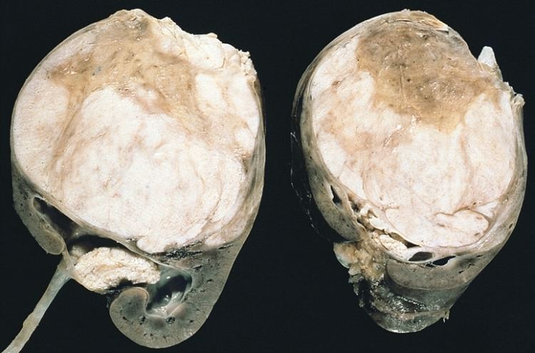
Most nephroblastomas are on one side of the body only and are found on both sides in less than 5% of cases, although patients with Denys-Drash syndrome mostly have bilateral or multiple tumors. They tend to be encapsulated and vascularized tumors that do not cross the midline of the abdomen. In cases of metastasis it is usually to the lung. A rupture of Wilms tumor puts the patient at risk of bleeding and peritoneal dissemination of the tumor. In such cases, surgical intervention by a surgeon who is experienced in the removal of such a fragile tumor is imperative.
Pathologically, a triphasic nephroblastoma comprises three elements:
Wilms tumor is a malignant tumor containing metanephric blastema, stromal and epithelial derivatives. Characteristic is the presence of abortive tubules and glomeruli surrounded by a spindled cell stroma. The stroma may include striated muscle, cartilage, bone, fat tissue, fibrous tissue. Dysfunction is caused when the tumor compresses the normal kidney parenchyma.
The mesenchymal component may include cells showing rhabdomyoid differentiation or malignancy (rhabdomyosarcomatous Wilms).
Wilms tumors may be separated into 2 prognostic groups based on pathologic characteristics:
Molecular biology
Mutations of the WT1 gene on chromosome 11p13 are observed in approximately 20% of Wilms tumors. At least half of the Wilms tumors with mutations in WT1 also carry mutations in CTNNB1, the gene encoding the proto-oncogene beta-catenin.
A gene on the X chromosome, WTX, is inactivated in up to 30% of Wilms tumor cases, according to research published in 2007.
Most cases do not have mutations in any of these genes.
Diagnosis
The first sign is normally a painless abdominal tumor that can be easily felt by the doctor. An ultrasound scan, computed tomography scan, or MRI scan is done first. A tumor biopsy is not typically performed due to the risk of creating fragments of cancer tissue and seeding the abdomen with malignant cells.
Staging
Staging is a standard way to describe the extent of spread of Wilms tumors, and to determine prognosis and treatments. Staging is based on anatomical findings and tumor cells pathology.
Stage I (43% of patients)
Stage I Wilms tumor, all of the following criteria must be met:
Stage II (23% of patients)
Stage II Wilms tumor, 1 or more of the following criteria must be met:
Stage III (20% of patients)
Stage III Wilms tumor, 1 or more of the following criteria must be met:
Stage IV (10% of patients)
Stage IV Wilms tumor is defined as the presence of hematogenous metastases (lung, liver, bone, or brain), or lymph node metastases outside the abdomenopelvic region.
Stage V (5% of patients)
Stage V Wilms tumor is defined as bilateral renal involvement at the time of initial diagnosis. Note: For patients with bilateral involvement, an attempt should be made to stage each side according to the above criteria (stage I to III) on the basis of extent of disease prior to biopsy.
Treatment/prognosis
The overall 5-year survival is estimated to be approximately 90%, but for individuals the prognosis is highly dependent on individual staging and treatment. Early removal tends to promote positive outcomes.
Tumor-specific loss-of-heterozygosity (LOH) for chromosomes 1p and 16q identifies a subset of Wilms tumor patients who have a significantly increased risk of relapse and death. LOH for these chromosomal regions can now be used as an independent prognostic factor together with disease stage to target intensity of treatment to risk of treatment failure. Genome-wide copy number and LOH status can be assessed with virtual karyotyping of tumor cells (fresh or paraffin-embedded).
Statistics may sometimes show more favorable outcomes for more aggressive stages than for less aggressive stages, which may be caused by more aggressive treatment and/or random variability in the study groups. Also, a stage V tumor is not necessarily worse than a stage IV tumor.
Epidemiology
People of African descent have the highest rates of Wilms' tumor. Most instances of cancer occur among children between 3 and 3.5 years old. A genetic predisposition to Wilms' Tumor in individuals with aniridia has been established, due to deletions in the p13 band on chromosome 11.
History
Dr. Sidney Farber, founder of Dana-Farber Cancer Institute, and his colleagues achieved the first remissions in Wilms tumor in the 1950s. By employing the antibiotic actinomycin D in addition to surgery and radiation therapy, they boosted cure rates from 40 to 89 percent...