
Symbol DctP Pfam clan CL0177 TCDB 2.A.56 | Pfam PF03480 InterPro IPR018389 Pfam structures | |
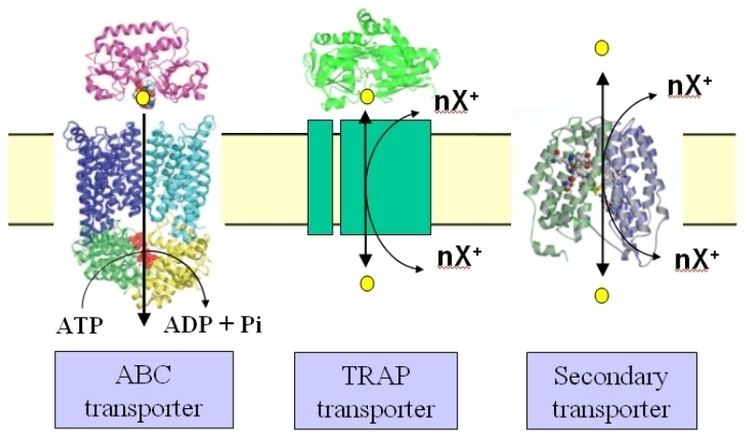 | ||
Tripartite ATP-independent periplasmic transporters (TRAP transporters) are a large family of solute transporters found in bacteria and archaea, but not in eukaryotes, that appear to be specific for the uptake of organic acids. They are unique in that they utilize a substrate binding protein (SBP) in combination with a secondary transporter.
Contents
History
TRAP transporters were discovered in the laboratory of Prof. David J. Kelly at the University of Sheffield, UK. His group were working on the mechanism used by the photosynthetic bacterium Rhodobacter capsulatus to take up certain dicarboxylic acids. They characterised a binding protein component (DctP) of a transporter that recognized these compounds, which they assumed would form part of a typical ABC transporter, but when they sequenced the genes surrounding dctP they found two other genes encoding integral membrane proteins, dctQ and dctM, but no genes encoding components of an ABC transporter. They further showed that uptake of the same dicarboxylates was independent of ATP and that uptake required an electrochemical ion gradient, making this a unique binding protein-dependent secondary transporter.
Since these early studies, it has become clear that TRAP transporters are present in many bacteria and archaea, with many bacterial having multiple TRAP transporters, some having over 20 different systems.
Substrates
To date, most substrates for TRAP transporters contain a common feature which is that they are organic acids. This includes C4-dicarboxylates such as succinate, malate and fumarate, keto-acids such as pyruvate and alpha-ketobutyrate and the sugar acid, N-acetyl neuraminic acid (or sialic acid). Other substrates include the compatible solute ectoine and hydroxyectoine and pyroglutamate.
Composition
All known TRAP transporters contain 3 protein domains. These are the solute binding protein (the SBP), the small membrane protein domain and the large membrane protein domain. Following the nomenclature for the first characterized TRAP transporter, DctPQM, these subunits are usually named P, Q and M respectively. Around 10% of TRAP transporters have natural genetic fusions between the two membrane protein components, and in the one well studied example of this in the sialic acid specific TRAP transporter from Haemophilus influenzae the fused gene has been named siaQM. The large M subunit is predicted to have 12 transmembrane helices and the small Q subunit to have 4 transmembrane helices and the fused QM proteins are predicted to have 17 transmembrane helices.
Mechanism
By using an SBP, TRAP transporters share some similarity to ABC transporters in that the substrate for the transporter is initially recognized outside of the cytoplasmic membrane. In Gram-negative bacteria, the SBP is usually free in the periplasm and expressed at relatively high levels compared to the membrane domains. In Gram positive bacteria and archaea, the SBP is tethered to the cytoplasmic membrane. In both types of systems the SBP binds to substrate, usually with low micromolar affinity, which causes a significant conformation change in the protein, akin to a Venus flytrap closing. The trapped subtrate is then delivered to the membrane domains of the transporter, where the electrochemical ion gradient is somehow exploited to open the SBP, extract the substrate and catalyse its movement across the membrane. For the SiaPQM TRAP transporter which has been studied in a fully reconstituted in vitro form, uptake uses a Na+
gradient and not proton gradient to drive uptake. The SiaPQM systems also exhibits unique properties for a secondary transporter in that it cannot catalyse bidirectional transport as the SBP imposes that movement is only in the direction of uptake into the cell.
Substrate binding protein (SBP)
Following the first structure of a TRAP SBP in 2005, there are now over 10 different structures available. They all have very similar overall structures, with two globular domains linked by a hinge. The substrate binding site is formed by both the domains which enclose the substrate. A highly conserved arginine residue in the TRAP SBPs forms a salt bridge with a carboxylate group on the substrate, which is important for substrate recognition.
Membrane subunits
There are currently no structures for the membrane domains of any TRAP transporter. It is not even known which subunit(s) made a direct interaction with the SBP subunit during the transport cycle.