
ICD-9-CM 756.0 DiseasesDB 13267 | ICD-10 Q75.4 OMIM 154500 | |
 | ||
Synonyms Treacher Collins–Franceschetti syndrome, mandibulofacial dysostosis, Franceschetti-Zwalen-Klein syndrome | ||
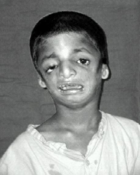
Treacher Collins syndrome (TCS) is an autosomal dominant congenital disorder characterized by craniofacial deformities, typically involving the ears, eyes, cheekbones, and jawbone. Those affected have normal intelligence. The typical physical features include downward-slanting eyes, micrognathia (a small lower jaw), conductive hearing loss, underdeveloped zygomatic bones, drooping part of the lateral lower eyelids, and malformed or absent ears, but they can vary dramatically between affected people. These physical features can cause problems breathing, hearing, and seeing.
Contents
- Signs and symptoms
- Genetics
- TCOF1
- Other mutations
- Genetic counselling
- Diagnosis
- Prenatal diagnosis
- Clinical findings
- Radiological findings
- Radiographs
- CT scan
- Differential diagnosis
- Treatment
- Hearing loss
- Epidemiology
- History
- Media portrayals
- References
TCS is most commonly caused by a mutation in the TCOF1 gene, but can also be caused by mutations in POLR1C or POLR1D, all of which are genes involved in the development of the pharyngeal arches and assembly of ribosomes, making Treacher Collins a ribosomopathy. It is diagnosed with a physical examination, although imaging can be used to investigate specific symptoms. Treacher Collins syndrome is not curable, but its symptoms can be managed with reconstructive surgery, hearing aids, and other assistive devices and practices.
TCS occurs in about one in 50,000 births in Europe. The syndrome is named after Edward Treacher Collins (1862–1932), an English surgeon and ophthalmologist, who described its essential traits in 1900.
Signs and symptoms
Symptoms in people with Treacher Collins syndrome vary. Some individuals are so mildly affected that they remain undiagnosed, while others have severe facial involvement and life-threatening airway compromise. Most of the features of TCS are symmetrical and are already recognisable at birth.
The most common symptom of Treacher Collins syndrome is hypoplasia (underdevelopment) of the mandible (lower jaw), also called micrognathia, and zygomatic bone, termed "malar hypoplasia". This can be accompanied by glossoptosis. Mandibular hypoplasia can result in a malocclusion, where the teeth and jaws do not line up properly, or in more severe cases, trouble breathing or swallowing. Underdevelopment of the zygomatic bone gives the cheeks a sunken appearance.
The external ear (pinna) is sometimes small, rotated, malformed, or absent entirely in people with TCS. Symmetric, bilateral narrowing (stenosis) or absence (atresia) of the external auditory canals is also described. In most cases, the ossicles and the middle ear cavity are misshapen. Inner ear malformations are rarely described. As a result of these abnormalities, a majority of the individuals with TCS have conductive hearing loss.
Most affected people also experience eye problems, including colobomata (notches) in the lower eyelids, partial or complete absence of eyelashes on the lower lid, downward angled eyelids, drooping of upper and lower eyelids (ptosis), and narrowing of the tear ducts (dacrostenosis). Vision loss can occur and is associated with strabismus, refractive errors, and anisometropia. It can also be caused by severely dry eyes, a consequence of lower eyelid abnormalities and frequent eye infections.
Although an abnormally shaped skull is not distinctive for Treacher Collins syndrome, brachycephaly with bitemporal narrowing is sometimes observed. Cleft palate is also common.
Dental anomalies are seen in 60% of affected people, including tooth agenesis (33%), discoloration (enamel opacities) (20%), malplacement of the maxillary first molars (13%), and wide spacing of the teeth. In some cases, dental anomalies in combination with mandible hypoplasia result in a malocclusion. This can lead to problems with food intake and the ability to close the mouth.
Less common features of TCS may add to an affected person's breathing problems, including sleep apnea. Choanal atresia or stenosis is a narrowing or absence of the choanae, the internal opening of the nasal passages. Pharyngeal hypoplasia, underdevelopment of the pharynx, can also narrow the airway.
Features related to TCS that are seen less frequently include nasal deformities, high-arched palate, macrostomia, preauricular hair displacement, cleft palate, hypertelorism, notched upper eyelid, and congenital heart defects.
The general public may associate facial deformity with developmental delay and intellectual disability, but more than 95% of people affected with TCS have normal intelligence. The psychological and social problems associated with facial deformity can affect quality of life in people with TCS.
Genetics
Mutations in TCOF1, POLR1C, or POLR1D genes can cause Treacher Collins syndrome. TCOF1 gene mutations are the most common cause of the disorder, accounting for 81 to 93% of all cases. POLR1C and POLR1D gene mutations cause an additional 2% of cases. In individuals without an identified mutation in one of these genes, the genetic cause of the condition is unknown. The TCOF1, POLR1C, and POLR1D genes code for proteins which play important roles in the early development of bones and other tissues of the face. Mutations in these genes reduce the production of rRNA, which may trigger the self-destruction (apoptosis) of certain cells involved in the development of facial bones and tissues. It is unclear why the effects of a reduction in rRNA are limited to facial development. Mutations in TCOF1 and POLR1D cause the autosomal dominant form of Treacher Collins, and mutations in POLR1C cause the autosomal recessive form.
TCOF1
TCOF1 is the only gene currently known to be associated with TCS, a mutation in this gene being found in 90-95% of the individuals with TCS. However, in some individuals with typical symptoms of TCS, mutations in TCOF1 have not been found. Investigation of the DNA has resulted in the identification of the kind of mutations found in TCOF1. The majority of mutations are small deletions or insertions, though splice site and missense mutations also have been identified.
Mutation analysis has unveiled more than 100 disease-causing mutations in TCOF1, which are mostly family-specific mutations. The only recurrent mutation accounts for about 17% of the cases.
TCOF1 is found on the 5th chromosome in the 5q32 region. It codes for a relatively simple nucleolar protein called treacle, that is thought to be involved in ribosome assembly. Mutations in TCOF1 lead to haploinsufficiency of the treacle protein. Haploinsufficiency occurs when a diploid organism has only one functional copy of a gene, because the other copy is inactivated by a mutation. The one normal copy of the gene does not produce enough protein, causing disease. Haploinsufficiency of the treacle protein leads to a depletion of the neural crest cell precursor, which leads to a reduced number of crest cells migrating to the first and second pharyngeal arches. These cells play an important role in the development of the craniofacial appearance, and loss of one copy of treacle affects the cells' ability to form the bones and tissues of the face.
Other mutations
POLR1C and POLR1D mutations are responsible for a minority of cases of Treacher Collins. POLR1C is found on chromosome 6 at position 6q21.2 and codes for a protein subunit of RNA polymerase I. POLR1D is found on chromosome 13 at position 13q12.2 and codes for a protein subunit of RNA polymerase III. Both of these polymerases are important for ribosome biogenesis.
Genetic counselling
TCS is inherited in an autosomal dominant manner and the penetrance of the affected gene is almost complete. Some recent investigations, though, described some rare cases in which the penetrance in TCS was not complete. Causes may be a variable expressivity, an incomplete penetrance or germline mosaicism. Only 40% of the mutations are inherited. The remaining 60% are a result of a de novo mutation, where a child has a new mutation in the responsible gene and did not inherit it from either parent. In the outcome of the disease, inter- and intrafamilial variability occurs. This suggests, when an affected child is born, it is important to investigate the parents to determine whether the affected gene is present, because the parent could have a mild form of the disease that has not been diagnosed. In this case, the risk of having another affected child is 50%. If the parents do not have the affected gene, the recurrence risk appears to be low. In following generations, the severity of the clinical symptoms increases.
Diagnosis
The diagnosis of Treacher Collins syndrome relies upon clinical and radiographic findings. Prenatal diagnosis cannot be guaranteed.
Prenatal diagnosis
Mutations in the main genes responsible for TCS can be detected with chorionic villus sampling or amniocentesis. Rare mutations may not be detected by these methods. Ultrasonography can be used to detect craniofacial abnormalities later in pregnancy, but may not detect milder cases.
Clinical findings
TCS is often first suspected with characteristic symptoms observed during a physical exam. However, the clinical presentation of TCS can resemble other diseases, making diagnosis difficult. The OMENS classification was developed as a comprehensive and stage-based approach to differentiate the diseases. This acronym describes five distinct dysmorphic manifestations, namely orbital asymmetry, mandibular hypoplasia, auricular deformity, nerve development, and soft-tissue disease.
Mandible
- 2A: glenoid fossa in anatomical acceptable position
- 2B: Temperomandibular joint inferiorly (TMJ), medially, anteriorly displaced, with severely hypoplastic condyle
Ear
Facial nerve
Soft tissue
Radiological findings
Radiologic manifestations can be used to confirm the diagnosis. Imaging evaluation techniques used include X-rays (radiographs), CT scans, MRI, and ultrasound.
Radiographs
A few techniques are used to confirm the diagnosis in TCS.
An orthopantomogram (OPG) is a panoramic dental X-ray of the upper and lower jaw. It shows a two-dimensional image from ear to ear. Particularly, OPG facilitates an accurate postoperative follow-up and monitoring of bone growth under a mono- or double-distractor treatment. Thereby, some TCS features could be seen on OPG, but better techniques are used to include the whole spectrum of TCS abnormalities instead of showing only the jaw abnormalities.
Another method of radiographic evaluation is taking an X-ray image of the whole head. The lateral cephalometric radiograph in TCS shows hypoplasia of the facial bones, like the malar bone, mandible, and the mastoid.
Finally, occipitomental radiographs are used to detect hypoplasia or discontinuity of the zygomatic arch.
CT scan
A temporal-bone CT using thin slices makes it possible to diagnose the degree of stenosis and atresia of the external auditory channel, the status of the middle ear cavity, the absent or dysplastic and rudimentary ossicles, or the inner ear abnormalities such as a deficient cochlea. Two- and three-dimensional CT reconstructions with VRT and bone and skin-surfacing are helpful for more accurate staging and the three-dimensional planning of mandibular and external ear reconstructive surgery.
Differential diagnosis
Other diseases have similar characteristics to Treacher Collins syndrome. In the differential diagnosis, one should consider the acrofacial dysostoses. The facial appearance resembles that of Treacher Collins syndrome, but additional limb abnormalities occur in those persons. Examples of these diseases are Nager syndrome and Miller syndrome. The oculoauriculovertebral spectrum should also be considered in the differential diagnosis. An example is hemifacial microsomia, which primarily affects development of the ear, mouth, and mandible. This anomaly may occur bilaterally. Another disease which belongs to this spectrum is Goldenhar syndrome, which includes vertebral abnormalities, epibulbar dermoids and facial deformities.
Treatment
The treatment of individuals with TCS may involve the intervention of professionals from multiple disciplines. The primary concerns are breathing and feeding, as a consequence of the hypoplasia of the mandibula and the obstruction of the hypopharynx by the tongue. Sometimes, they may require a tracheostomy to maintain an adequate airway, and a gastrostomy to assure an adequate caloric intake while protecting the airway. Corrective surgery of the face is performed at defined ages, depending on the developmental state.
An overview of the present guidelines:
- Type I (mild) and Type IIa (moderate) 13–16 years
- Type IIb (moderate to severe malformation) at skeletal maturity
- Type III (severe) 6–10 years
Hearing loss
Hearing loss in Treacher Collins syndrome is caused by deformed structures in the outer and middle ear. The hearing loss is generally bilateral with a conductive loss of about 50-70 dB. Even in cases with normal auricles and open external auditory canals, the ossicular chain is often malformed.
Attempts to surgically reconstruct the external auditory canal and improve hearing in children with TCS have not yielded positive results.
Auditory rehabilitation with bone-anchored hearing aids (BAHAs) or a conventional bone conduction aid has proven preferable to surgical reconstruction.
Epidemiology
TCS occurs in about one in 50,000 births in Europe. Worldwide, it is estimated to occur in one in 10,000 to one in 50,000 births.
History
The syndrome is named after Edward Treacher Collins (1862–1932), the English surgeon and ophthalmologist who described its essential traits in 1900. In 1949, Adolphe Franceschetti and David Klein described the same condition on their own observations as mandibulofacial dysostosis. The term mandibulofacial dysostosis is used to describe the clinical features.
Media portrayals
A July 1977 New York Times article that was reprinted in numerous newspapers nationwide over the ensuing weeks brought this malady to many people's attention for the first time.
The disorder was featured on the show Nip/Tuck, in the episode "Blu Mondae".
TLC's Born Without a Face features Juliana Wetmore, who was born with the most severe case in medical history of this syndrome and is missing 30%–40% of the bones in her face.
In 2010, BBC Three documentary Love Me, Love My Face covered the case of a man, Jono Lancaster, with the condition. In 2011, BBC Three returned to Jono to cover his and his partner Laura's quest to start a family, in So What If My Baby Is Born Like Me?, which first aired as part of a BBC Three season of programmes on parenting. The first film was replayed on BBC One shortly ahead of the second film's initial BBC Three broadcast. Lancaster's third BBC Three film, Finding My Family on Facebook, which looked at adoption, aired in 2011.
In Wonder, the children's novel, the main character is a child who has Treacher Collins Syndrome.