
TA A14.2.01.160 | Dorlands/Elsevier n_05/12566088 FMA 6239 | |
 | ||
Latin nervus laryngeus superior | ||
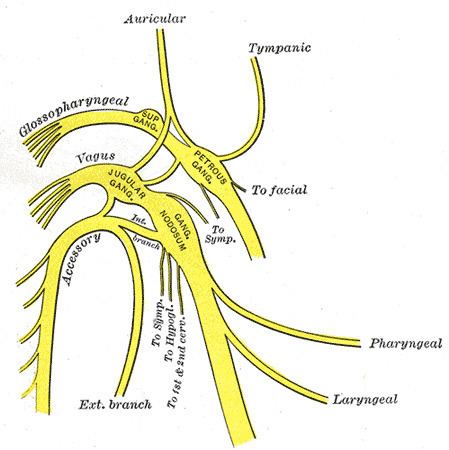
The superior laryngeal nerve is a branch of the vagus nerve. It arises from the middle of the inferior ganglion of vagus nerve and in its course receives a branch from the superior cervical ganglion of the sympathetic nervous system.
Contents
All intrinsic laryngeal muscles except the cricothyroids are innervated by the recurrent laryngeal nerve. The cricothyroid muscles are innervated by the superior laryngeal nerve.
The recurrent laryngeal nerve gets its name from the fact that it loops below the aorta on its way to the intrinsic muscles of the larynx. The left recurrent laryngeal nerve passes under and around the aorta on its way to the larynx, whereas the right recurrent laryngeal nerve passes under and around the subclavian artery. Because the aorta is inferior to the subclavian artery, the left recurrent laryngeal nerve is a bit longer than the right recurrent laryngeal nerve. However, there is no discernible effect on the timing of neural impulses to the muscles these two nerves serve. By comparison with the recurrent laryngeal nerves, the superior laryngeal nerve takes a more direct route on the way to the cricothyroid muscles.
Structure
The superior laryngeal nerve descends, by the side of the pharynx, behind the internal carotid artery, and divides into two branches —the external laryngeal nerve and the internal laryngeal nerve.
The external laryngeal nerve is the smaller, external branch. It descends on the larynx, beneath the sternothyroid muscle, to supply the cricothyroid muscle. The external branch functions to tense the vocal cords by activating the cricothyroid muscle, increasing pitch. The external laryngeal nerve gives branches to pharyngeal plexus and the superior portion of the inferior pharyngeal constrictor, and communicates with the superior cardiac nerve behind the common carotid artery.
The internal laryngeal nerve is the internal branch. It descends to the thyrohyoid membrane, piercing it in company with the superior laryngeal artery, and is distributed to the mucous membrane of the larynx. Of these sensory branches, some are distributed to the epiglottis, the base of the tongue, and the epiglottic glands; others pass posteriorly, in the aryepiglottic fold, to supply the mucous membrane surrounding the entrance of the larynx, and the mucous lining of the larynx as far down as the vocal folds.
A filament descends beneath the mucous membrane on the inner surface of the thyroid cartilage and joins the recurrent nerve. Above the vocal folds the sensory innervation of the larynx is via the internal laryngeal nerve. Below the vocal folds it is by way of branches of the recurrent laryngeal nerve. The vocal fold itself receives dual innervation from both nerves.
Clinical significance
A superior laryngeal nerve palsy changes the pitch of the voice and causes an inability to make explosive sounds due to paralysis of the cricothyroid muscle. If no recovery is evident three months after the palsy initially presents, the damage is most likely to be permanent. A bilateral palsy presents as a tiring and hoarse voice. Damage to the superior laryngeal nerve leaves the vocal cord abducted and poses an aspiration risk. It can be injured in surgery involving the removal of the thyroid gland (thyroidectomy).
The external branch is susceptible to damage during thyroidectomy or cricothyrotomy, as it lies immediately deep to the superior thyroid artery. The ability to produce pitched sounds is then impaired along with easy voice fatigability, (usually mono-toned voice).
Irritation of the internal laryngeal nerve results in uncontrolled coughing - usually as a result of food or water in the laryngopharynx.