
Order Fusobacteriales | Family Leptotrichiaceae Higher classification Streptobacillus | |
 | ||
Scientific name Streptobacillus moniliformis Similar Streptobacillus, Bacteria, Spirillum minus, Spirillum, Eikenella corrodens | ||
Pronounce streptobacillus moniliformis speakmedical
Streptobacillus moniliformis is a non-motile, Gram-negative rod-shaped bacterium that is a member of the family Leptotrichiaceae. The genome of S. moniliformis is one of two completed sequences of the order Fusobacteriales. Its name comes the Greek word streptos for "curved" or "twisted", and the Latin words bacillus meaning "small rod" and moniliformis for "necklace". S. moniliformis is microaerophilic, requiring less oxygen than is present in the atmosphere for its growth.
Contents
- Pronounce streptobacillus moniliformis speakmedical
- History
- Microbiology
- Morphology
- Taxonomy
- Genomics
- Isolation and identification
- Rat bite fever and Haverhill fever
- References

History
S. moniliformis was first isolated from a rat-bitten man in 1914 by German microbiologist H. Schottmüller, who described it as Streptothrix muris ratti. In the United States during the year 1916, S. moniliformis was determined to be the causative source of rat-bite fever.
Microbiology
Some isolates of S. moniliformis have been collected from the upper respiratory tract of domestic and wild rats. Two known variants of S. moniliformis have been identified. The bacillary type is pathogenic. In contrast, the spontaneously occurring L form, which lacks a cell wall and whose colonies grow in a "fried egg" formation, is non-pathogenic.
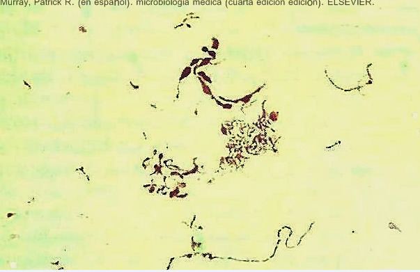
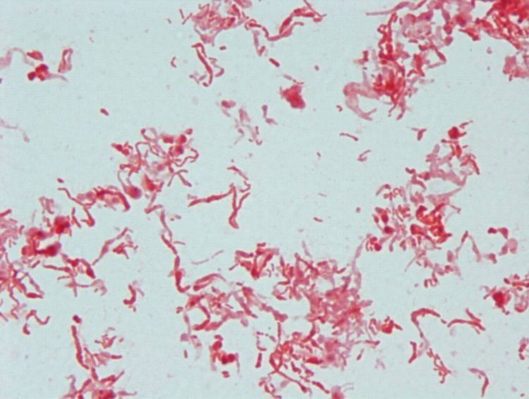
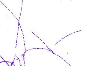
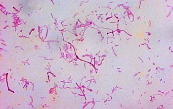
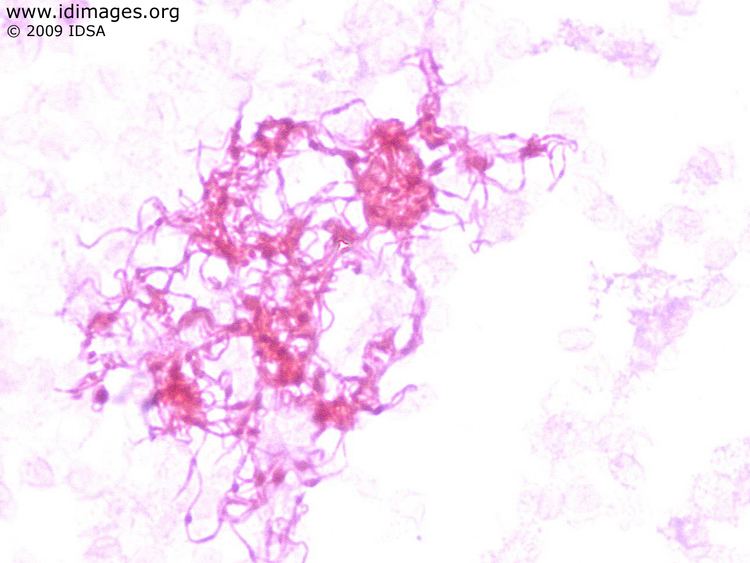
Morphology
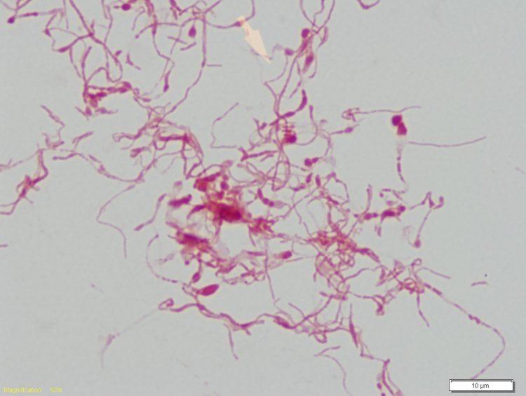
S. moniliformis has frequently been observed in the form of filamentous, non-branching chains and is highly pleomorphic. For example, the normally straight rod can develop lateral bulbar swellings. The bacteria vary in size from 0.1 to 0.5 μm by 2.0 to 5.0 μm, and can potentially grow up to 10 to 15 μm, with long, curved segments from 100 to 150 μm.
Taxonomy
S. moniliformis was previously classified under the Fusobacteriaceae family. It was later regrouped with three other genera including Sebaldella, Sneathia, and Leptotrichia. These four genera were classified under the family Leptotrichiaceae following comparative analyses of the 16S ribosomal RNA gene sequences and 16S-23S rDNA internal transcribed spacer sequences among members of the phylum Fusobacteria. Identification of conserved signature indels unique to Fusobacteria and its primary clades as well as phylogenetic analysis of members of Fusobacteria based on concatenated sequences of 17 conserved proteins further support the distinction between the two families.
S. moniliformis was formerly classified as the only member of the Streptobacillus genus. However, Streptobacillus strains HKU33T and HKU34 were isolated in Hong Kong in September 2014. Streptobacillus HKU33T was found in pus isolated from the abscess of a 38-year-old patient with quinsy and HKU34 from the elbow joint fluid of a 64-year-old patient with septic arthritis. Following analysis of the 16S rRNA gene sequences found in members of Leptotrichiaceae and partial sequences of the recA, groEl, and gyrB genes present in both isolates, the two strains were taxonomically grouped under the novel species Streptobacillus hongkongensis sp. nov.
Genomics
The U.S. Department of Energy's Joint Genome Institute (DOE JGI) sequenced the complete genome of S. moniliformis DSM 12112. It is made up of one circular chromosome of 1,673,280 base pairs as determined from a combination of Sanger and 454 sequencing. The mol% of guanine and cytosine in the DNA is 26.3% with 1,511 protein coding genes out of the 1,566 genes predicted. These low G+C values were previously only seen in members of the order Mycoplasmatales, which includes the genus Mycoplasma, indicating a relationship between Mycoplasma and S. moniliformis. However, 16S rRNA gene analysis showed this relation to be incorrect. S. moniliformis also has a single circular plasmid pSMON01 that is 10,702 base pairs long with 1,511 protein coding genes.
Isolation and identification
Isolation and identification is challenging because growth of the bacteria is inhibited by sodium polyanethol sulfonate, an anticoagulant present in most commercial aerobic blood culture bottles that are used as enriched growth media. Trypticase soy agar or broth containing 10-20% blood, serum, or ascites fluid, as well as anaerobic culture bottles and resin bead culture systems can be used for growth because sodium polyanethol sulfonate is not normally present. Samples from the inner ear of rats have been used to inoculate 5% sheep blood agar, which ranges in pH from 7.2 to 7.6. The samples were incubated at 37 °C, the same as the average temperature of the human body. It typically takes 2 to 3 days for colonies to appear, however it may take as long as 7. Colonies appear round, convex, smooth, shiny, and greyish in color on solid agar but, in liquid media, colonies appear as "cotton balls." Once they have been cultured, confirmation of their identity can be made using conventional biochemical analyses, such as tests for the production of oxidase, catalase, indole, nitrate, as well as carbohydrate fermentation. S. moniliformis can be biochemically differentiated by similar bacteria by their negative production of indole, catalase, and oxidase, and reduction of nitrate to nitrite. S. moniliformis has been shown to be sensitive to penicillin, cephalosporins, macrolides, tetracyclines and other broad spectrum antibiotics.
Rat bite fever and Haverhill fever
In the U.S., rat bite fever is primarily caused by transmission of S. moniliformis from the bite of a rat. However, approximately 30% of patients diagnosed with rat bite fever do not recall being scratched or bitten by an infected animal. Transmission of the bacterium is also known to occur via consumption of infected water, close contact with, or handling of rats. Haverhill fever, named after the 1926 outbreak of the disease in Haverhill, Massachusetts, is a form of rat bite fever that can result from ingesting food contaminated with S. moniliformis. In 1986 at a boarding school in the United Kingdom, another outbreak of Haverhill fever was reported. Some 304 people were reported to have been afflicted. Infection was suspected to have resulted from the consumption of either unpasteurized milk or water contaminated with rat feces. Infected individuals described symptoms including a sudden development of vomiting, severe headache, and cold sweats with a high fever. Parker and Hudson first isolated the cause of this outbreak, which they named Haverhilia multiformis. This organism was later matched to S. moniliformis after further research.
Symptoms of rate bite fever include the abrupt onset of fever ranging from 38.0 °C to 41 °C. Approximately 75% of infected individuals develop a rash in addition to hemorrhaging vesicles. Both the rash and vesicles are usually located on the hands and feet, although the rash has been known to spread to other parts of the body.
The microaerophilic nature of S. moniliformis makes identification difficult. PCR testing is being utilized more for its identification. However, there is still a 13% mortality rate for untreated cases. Immunocompromised individuals, such as HIV-positive individuals, are more at risk of death from this disease. Lab personnel and pet store workers, who work closely with animals on a daily basis, also have an increased risk of infection.
Although S. moniliformis is believed to be part of the commensal bacteria of the respiratory tract of rats, rats have occasionally shown signs of the disease. Antibiotics used to treat infection may cause the formation of the L-form, which persists in the body although this form is not pathogenic.