
 | ||
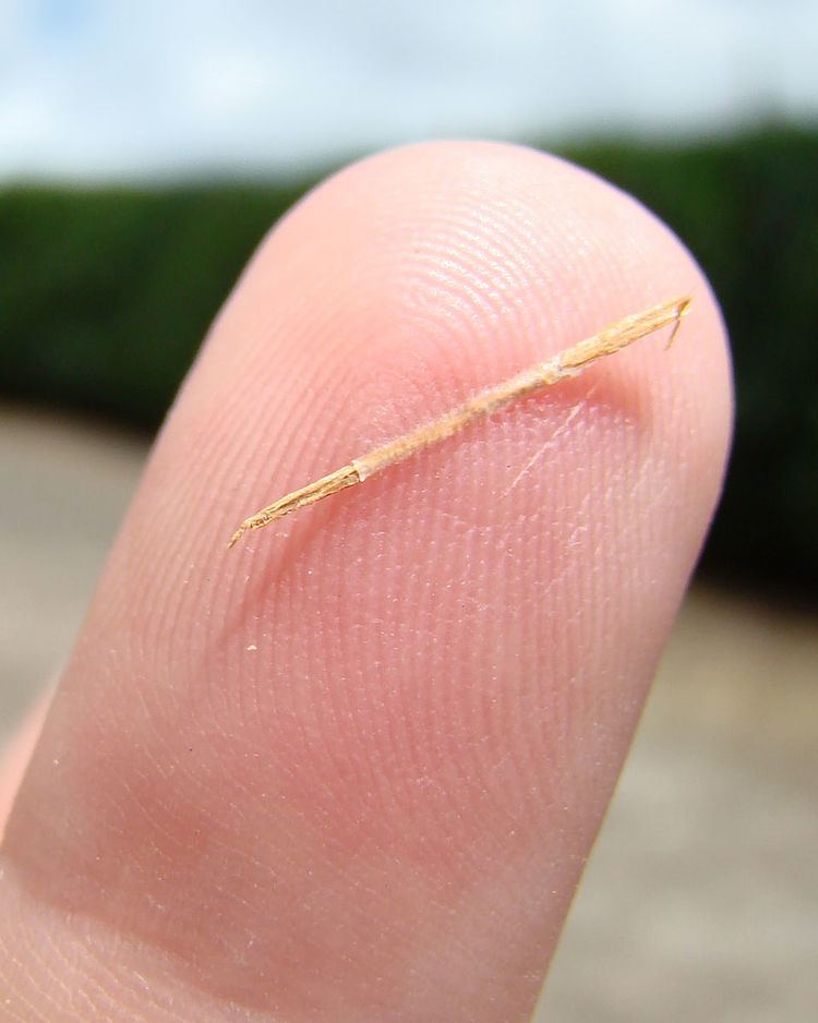
A splinter is a fragment of a larger object (especially wood), or a foreign body that penetrates or is purposely injected into a body. The foreign body must be lodged inside tissue to be considered a splinter. Splinters may cause initial pain through ripping of flesh and muscle, infection through bacteria on the foreign object, and severe internal damage through migration to vital organs or bone over time.
Contents
- Contracting a splinter
- Types
- Specific details of some splinters
- Detection
- Signs of a hidden foreign body
- Removal
- Infection
- References
Splinters commonly consist of wood, but there are many other types. According to the American Academy of Family Physicians (AAFP), common types of splinters are glass, plastic, metal, and spines of animals.
Contracting a splinter
Generally, a splinter causes an initial feeling of pain as the sharp object makes its initial penetration through the body. Through this penetration, the object cuts through the cutaneous layer of the skin, and settles in the subcutaneous layer of the skin, and can even penetrate further down, breaking the sub-cutaneous layer, settling in muscle tissue, or even the bone. Some splinters will remain in place, but most will continue to migrate through the body, further damaging their surroundings.
Types
According to the AAFP, some of the most common foreign bodies contracted by people fall into two official classes: Biological splinters, and Nonbiological Splinters. Within the class of biological splinters such foreign bodies include bone, fish spines, teeth, and wood. Within the nonbiological class some of the most common splinters contracted by humans are glass, metal, aluminum, fishhooks, pencil graphite, and plastic.
Rarely, one may become infected with splinters from more unusual sources. Common cases of exotic foreign bodies include sea urchins, insect stings, stingray spines, and even grenade shrapnel.
Specific details of some splinters
Detection
Splinters are often first detected by the person with the splinter in their body. There are many signs that a splinter has entered one’s body.
Signs of a hidden foreign body
Many times, if the presence in a body is not known, but only effects are felt, modern technologies will be employed. Depending on the type of foreign body the patient is infected with modern detectors can range from Computed Tomography, Radiography, and Ultrasonography. If manual detection and localization fail, such modern technologies may be utilized accordingly:
Removal
There are many medical techniques to remove splinters safely. Common medical techniques include the Elliptical Technique and the String Technique. In the elliptical technique the surrounding area of the splinter is sliced in an elliptical formation. From there the flesh in the elliptical area is cut (in the shape of an upside-down cone) and the whole chunk of flesh, containing the splinter is removed. The Needle Cover Technique is limited to fishhook removal. A string is looped around the base of the hook, and as the hook is pressed further into the skin, the string is pulled, allowing the barbs to be unhooked from muscle and follow the path of the rest of the hook out of the body without snagging any additional flesh.
Infection
Infection is usually determined by the duration of time that the foreign object remains lodged in the human body. Objects that have included poison, deep penetration, dirt, or bite injuries generally result in a shorter time until infection is notable. According to the AAFP patients that are older, or have diabetes, or have wounds that are longer, wider, more jagged or deeper, have a much higher risk of infection. Simply the easiest way to avoid infection is to completely remove the splinters or foreign body as soon as possible. Though infection is generally the largest complication encountered with splinters, ranging from 1.1 to 12 percent presence, the use of antibiotics in non-bite cases is generally deemed unnecessary by the medical community. Though cases are rare, infection of foreign body wounds can result in cases of tetanus.
One case of tetanus contraction through a splinter was seen in Ohio, 1993. An 80-year-old woman was presented to an ED with dysphagia and a stiff jaw. Not long after a preliminary checkup, a wood splinter was found to have been lodged in her chin for approximately 1 week; the area was erythematous with active purulent drainage. The woman was diagnosed with tetanus, admitted to the hospital, and begun on a regimen of 3,000 units of tetanus immune globulin, tetanus toxoid, and intravenous clindamycin. Despite aggressive treatment, including assisted mechanical ventilation, the patient succumbed to the effects of her primary infection and died 15 days later. The woman had no history of previous tetanus vaccinations despite previous care for a wound and on-going medical attention for hypertension.