
Specialty rheumatology ICD-9-CM 719.65 eMedicine sports/123 | ICD-10 M76.1 DiseasesDB 32612 | |
 | ||
Snapping hip syndrome (also referred to as coxa saltans, iliopsoas tendinitis, or dancer's hip) is a medical condition characterized by a snapping sensation felt when the hip is flexed and extended. This may be accompanied by an audible snapping or popping noise and pain or discomfort. Pain often decreases with rest and diminished activity. Snapping hip syndrome is classified by location of the snapping, either extra-articular or intra-articular.
Contents
- Symptoms
- Extra articular
- Intra articular
- Causes of injury
- Extra articular snapping hip syndrome
- Intra articular snapping hip syndrome
- Treatment
- Self treatment
- Diagnostic imaging
- Injection based treatments
- Surgical treatment
- Rehabilitation
- Physical therapy or athletic training therapy and rehabilitation
- Images
- References
Symptoms
In some cases, an audible snapping or popping noise as the tendon at the hip flexor crease moves from flexion (knee toward waist) to extension (knee down and hip joint straightened). After extended exercise pain or discomfort may be present caused by inflammation of the iliopsoas bursae. Pain often decreases with rest and diminished activity. Symptoms usually last months or years without treatment and can be very painful.
Extra-articular
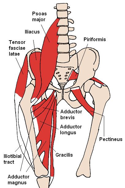
The more common lateral extra articular type of snapping hip syndrome occurs when the iliotibial band, tensor fasciae latae, or gluteus medius tendon slides back and forth across the greater trochanter. This normal action becomes a snapping hip syndrome when one of these connective tissue bands thickens and catches with motion. The underlying bursa may also become inflamed, causing a painful external snapping hip syndrome.
Less commonly, the iliopsoas tendon catches on the anterior inferior iliac spine (AIIS), the lesser trochanter, or the iliopectineal ridge during hip extension, as the tendon moves from an anterior lateral (front, side) to a posterior medial (back, middle) position. With overuse, the resultant friction may eventually cause painful symptoms, resulting in muscle trauma, bursitis, or inflammation in the area.
Intra-articular
Because the iliopsoas or hip flexor crosses directly over the anterior suprior labrum of the hip, an intra-articular hip derangement (i.e. labral tears, hip impingement, loose bodies) can lead to an effusion that subsequently produces internal snapping hip symptoms.
Causes of injury
The cause of snapping hip syndrome is not well understood, and confusion exists within the medical community regarding causation. Athletes appear to be at an enhanced risk for snapping hip syndrome due to repetitive and physically demanding movements. In athletes such as ballet dancers, gymnasts, horse riders, track and field athletes and soccer players, military training, or any vigorous exerciser, repeated hip flexion leads to injury. In excessive weightlifting or running, the cause is usually attributed to extreme thickening of the tendons in the hip region. Snapping hip syndrome most often occurs in people who are 15 to 40 years old.
Extra-articular snapping hip syndrome
Extra-articular snapping hip syndrome is commonly associated with leg length difference (usually the long side is symptomatic), tightness in the iliotibial band (ITB) on the involved side, weakness in hip abductors and external rotators, poor lumbopelvic stability and abnormal foot mechanics (overpronation). Popping occurs when the thickened posterior aspect of the ITB or the anterior gluteus maximus rubs over the greater trochanter as the hip is extended.
Intra-articular snapping hip syndrome
Similar causes as extra-articular snapping hip syndrome but often with an underlying mechanical problem in the lower extremity. The pain associated with internal variety tends to be more intense and therefore more debilitating than the external variety. Intra-articular snapping hip syndrome is often indicative of injury such as a torn acetabular labrum, recurrent hip subluxation, ligamentum teres tears, loose bodies, articular cartilage damage, or synovial chondromatosis (cartilage formations in the synovial membrane of the joint).
Treatment
This condition is usually curable with appropriate treatment, or sometimes it heals spontaneously. If it is painless, there is little cause for concern.
Correcting any contributing biomechanical abnormalities and stretching tightened muscles, such as the iliopsoas muscle or iliotibial band, is the goal of treatment to prevent recurrence.
Referral to an appropriate professional for an accurate diagnosis is necessary if self treatment is not successful or the injury is interfering with normal activities. Medical treatment of the condition requires determination of the underlying pathology and tailoring therapy to the cause. The examiner may check muscle-tendon length and strength, perform joint mobility testing, and palpate the affected hip over the greater trochanter for lateral symptoms during an activity such as walking.
Self-treatment
A self-treatment recommended by the U.S. Army for a soft tissue injury of the iliopsoas muscle treatment, like for other soft tissue injuries, is a HI-RICE (Hydration, Ibuprofen, Rest, Ice, Compression, Elevation) regimen lasting for at least 48 to 72 hours after the onset of pain. "Rest" includes such commonsense prescriptions as avoiding running or hiking (especially on hills), and avoiding exercises such as jumping jacks, sit-ups or leg lifts/flutter kicks.
Stretching of the tight structures (piriformis, hip abductor, and hip flexor muscle) may alleviate the symptoms. The involved muscle is stretched (for 30 seconds), repeated three times separated by 30 second to 1 minute rest periods, in sets performed two times daily for six to eight weeks. This should allow the soldier to progress back into jogging until symptoms disappear.
Diagnostic imaging
Injection based treatments
Injections are usually focused on the iliopsoas bursa. Corticosteroid injections are common, but usually only last weeks to months. In addition, corticosteroid side effects can include weight gain, weakening of the surrounding tissues, and more. Cellular based therapy may have a role in future injection based treatments, though there is no current research proving the effectiveness of these therapies.
Surgical treatment
If medicine or physical therapy is ineffective or abnormal structures are found, surgery may be recommended.
Surgical treatment is rarely necessary unless intra-articular pathology is present. In patients with persistently painful iliopsoas symptoms surgical release of the contracted iliopsoas tendon has been used since 1984. Iliopsoas and iliotibial band lengthening can be done arthroscopically. Postop, these patients will usually undergo extensive physical therapy; regaining full strength may take up to 9–12 months.
Rehabilitation
Patients may require intermittent NSAID therapy or simple analgesics as they progress in activities. If persistent pain caused by bursitis continues a corticosteroid injection may be beneficial.
Physical therapy or athletic training therapy and rehabilitation
Both active and passive stretching exercises that include hip and knee extension should be the focus of the program. Stretching the hip into extension and limiting excessive knee flexion avoids placing the rectus femoris in a position of passive insufficiency, thereby maximizing the stretch to the iliopsoas tendon. Strengthening exercises for the hip flexors may also be an appropriate component of the program. A non-steroidal anti-inflammatory drug regimen as well as activity modification or activity progression (or both) may be used. Once symptoms have decreased a maintenance program of stretching and strengthening can be initiated. Light aerobic activity (warmup) followed by stretching and strengthening of the proper hamstring, hip flexors, and iliotibial band length is important for reducing recurrences.
Conservative measures may resolve the problem in 6 to 8 weeks.