
DiseasesDB 12185 eMedicine article/413810 | MedlinePlus 000972 | |
 | ||
Slipped capital femoral epiphysis (SCFE or skiffy, slipped upper femoral epiphysis, SUFE or souffy, coxa vara adolescentium) is a medical term referring to a fracture through the growth plate (physis), which results in slippage of the overlying end of the femur (epiphysis). Normally, the head of the femur, called the capital, should sit squarely on the femoral neck. Abnormal movement along the growth plate results in the slip. The femoral epiphysis remains in the acetabulum (hip socket), while the metaphysis (end of the femur) move in an anterior direction with external rotation.
Contents
- Classification
- Signs and symptoms
- Cause
- Pathophysiology
- Diagnosis
- Treatment
- Complications
- Epidemiology
- References
SCFE is the most common hip disorder in adolescence. SCFEs usually cause groin pain on the affected side, but sometimes cause knee or thigh pain. One in five cases involve both hips, resulting in pain on both sides of the body. SCFEs often occur in obese adolescent males, especially young Black males, although it also affects females. Symptoms include the gradual, progressive onset of thigh or knee pain with a painful limp. Hip motion will be limited, particularly internal rotation. Running and other strenuous activity on legs will also cause the hips to abnormally move due to the condition and can potentially worsen the pain. Stretching is very limited.
Classification
Signs and symptoms
Usually, a SCFE causes groin pain, but it may cause pain in only the thigh or knee, because the pain may be referred along the distribution of the obturator nerve. The pain may occur on both sides of the body (bilaterally), as up to 40 percent of cases involve slippage on both sides. After a first SCFE, when a second SCFE occurs on the other side, it typically happens within one year after the first SCFE. About 20 percent of all cases include a SCFE on both sides at the time of presentation.
Signs of a SCFE include a waddling gait, decreased range of motion. Often the range of motion in the hip is restricted in internal rotation, abduction, and flexion. A person with a SCFE may prefer to hold their hip in flexion and external rotation.
Cause
In general, SCFE is caused by increased force applied across the ephysis, or a decrease in the resistance within the physis to shearing. No single cause accounts for SCFEs, as several factors play a role in the development of a SCFE, particularly mechanical and endocrine (hormone-related) factors. Mechanical risk factors include obesity, coxa profunda, femoral or acetabular retroversion. Obesity is the most significant risk factor. In 65 percent of cases of SCFE, the person is over the 95th percentile for weight. Common misconception is heredity. Majority of cause is due to being overweight. Endocrine diseases also contribute, such as hypothyroidism, hypopituitarism, and renal osteodystrophy.
Pathophysiology
SCFE is a Salter-Harris type 1 fracture through the proximal femoral physis. Stress around the hip causes a shear force to be applied at the growth plate. While trauma has a role in the manifestation of the fracture, an intrinsic weakness in the physeal cartilage also is present. The almost exclusive incidence of SCFE during the adolescent growth spurt indicates a hormonal role. Obesity is another key predisposing factor in the development of SCFE.
The fracture occurs at the hypertrophic zone of the physeal cartilage. Stress on the hip causes the epiphysis to move posteriorly and medially. By convention, position and alignment in SCFE is described by referring to the relationship of the proximal fragment (capital femoral epiphysis) to the normal distal fragment (femoral neck). Because the physis has yet to close, the blood supply to the epiphysis still should be derived from the femoral neck; however, this late in childhood, the supply is tenuous and frequently lost after the fracture occurs. Manipulation of the fracture frequently results in osteonecrosis and the acute loss of articular cartilage (chondrolysis) because of the tenuous nature of the blood supply.
Diagnosis
The diagnosis is a combination of clinical suspicion plus radiological investigation. Children with a SCFE experience a decrease in their range of motion, and are often unable to complete hip flexion or fully rotate the hip inward. 20-50% of SCFE are missed or misdiagnosed on their first presentation to a medical facility. SCFEs may be initially overlooked, because the first symptom is knee pain, referred from the hip. The knee is investigated and found to be normal.
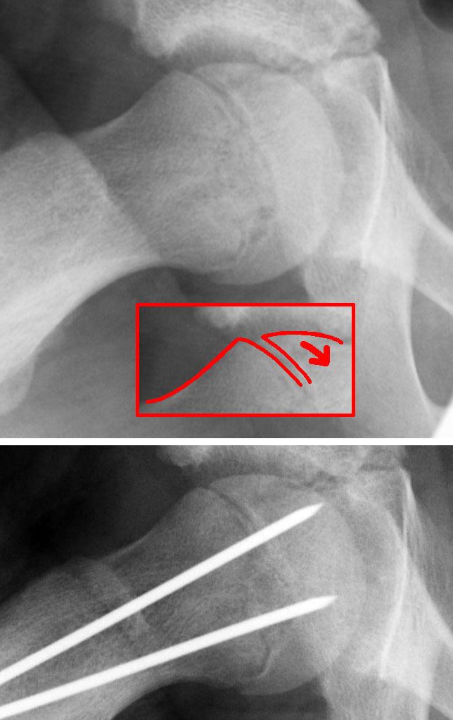
The diagnosis requires x-rays of the pelvis, with anteriorposterior (AP) and frog-leg lateral views. The appearance of the head of the femur in relation to the shaft likens that of a "melting ice cream cone", visible with Klein's line. The severity of the disease can be measured using the Southwick angle.
Treatment
The disease can be treated with external in-situ pinning or open reduction and pinning. Consultation with an orthopaedic surgeon is necessary to repair this problem. Pinning the unaffected side prophylactically is not recommended for most patients, but may be appropriate if a second SCFE is very likely.
Once SCFE is suspected, the patient should be non-weight bearing and remain on strict bed rest. In severe cases, after enough rest the patient may require physical therapy to regain strength and movement back to the leg. A SCFE is an orthopaedic emergency, as further slippage may result in occlusion of the blood supply and avascular necrosis (risk of 25 percent). Almost all cases require surgery, which usually involves the placement of one or two pins into the femoral head to prevent further slippage. The recommended screw placement is in the center of the epiphysis and perpendicular to the physis. Chances of a slippage occurring in the other hip are 20 percent within 18 months of diagnosis of the first slippage and consequently the opposite unaffected femur may also require pinning.
The risk of reducing this fracture includes the disruption of the blood supply to the bone. It has been shown in the past that attempts to correct the slippage by moving the head back into its correct position can cause the bone to die. Therefore the head of the femur is usually pinned 'as is'. A small incision is made in the outer side of the upper thigh and metal pins are placed through the femoral neck and into the head of the femur. A dressing covers the wound.
Complications
Failure to treat a SCFE may lead to: death of bone tissue in the femoral head (avascular necrosis), degenerative hip disease (hip osteoarthritis), gait abnormalities and chronic pain. SCFE is associated with a greater risk of arthritis of the hip joint later in life. 17-47 percent of acute cases of SCFE lead to the death of bone tissue (osteonecrosis) effects.
Epidemiology
SCFE affects approximately 1-10 per 100,000 children. The incidence varies by geographic location, season of the year, and ethnicity. In eastern Japan, the incidence is 0.2 per 100,000 and in the northeastern U.S. it is about 10 per 100,000. Africans and Polynesians have higher rates of SCFE.
SCFEs are most common in adolescents 11–15 years of age, and affects boys more frequently than girls (male 2:1 female). It is strongly linked to obesity, and weight loss may decrease the risk. Other risk factors include: family history, endocrine disorders, radiation / chemotherapy, and mild trauma.
The left hip is more often affected than the right. Over half of cases may have involvement on both sides (bilateral).