
 | ||
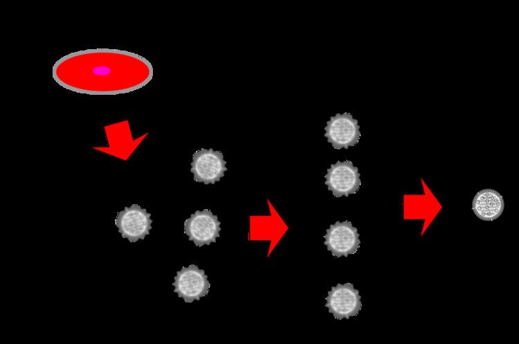
Single particle analysis is a group of related computerized image processing techniques used to analyze images from transmission electron microscopy (TEM). These methods were developed to improve and extend the information obtainable from TEM images of particulate samples, typically proteins or other large biological entities such as viruses. Individual images of stained or unstained particles are very noisy, and so hard to interpret. Combining several digitized images of similar particles together gives an image with stronger and more easily interpretable features. An extension of this technique uses single particle methods to build up a three-dimensional reconstruction of the particle. Using cryo-electron microscopy it has become possible to generate reconstructions with sub-nanometer resolution and near-atomic resolution first in the case of highly symmetric viruses, and now in smaller, asymmetric proteins as well.
Contents
Techniques
Single particle analysis can be done on both negatively stained and vitreous ice-embedded cryo-EM samples. Single particle analysis methods are, in general, reliant on the sample being homogeneous, although techniques for dealing with conformational heterogeneity are being developed.
Images (micrographs) collected on film are digitized using high-quality scanners, although increasingly electron microscopes have built-in CCD detectors coupled to a phosphorescent layer. The image processing is carried out using specialized software programs (for instance ), often run on multi-processor computer clusters. Depending on the sample or the desired results, various steps of two- or three-dimensional processing can be done.
Alignment and classification
Biological samples, and especially samples embedded in thin vitreous ice, are highly radiation sensitive, thus only low electron doses can be used to image the sample. This low dose, as well as variations in the metal stain used (if used) means images have high noise relative to the signal given by the particle being observed. By aligning several similar images to each other so they are in register and then averaging them, an image with higher signal to noise ratio can be obtained. As the noise is mostly randomly distributed and the underlying image features constant, by averaging the intensity of each pixel over several images only the constant features are reinforced. Typically, the optimal alignment (a translation and an in-plane rotation) to map one image onto another is calculated by cross-correlation.
However, a micrograph often contains particles in multiple different orientations and/or conformations, and so to get more representative image averages, a method is required to group similar particle images together into multiple sets. This is normally carried out using one of several data analysis and image classification algorithms, such as multi-variate statistical analysis and hierarchical ascendant classification, or K-means classification.
Often data sets of tens of thousands of particle images are used, and to reach an optimal solution an iterative procedure of alignment and classification is used, whereby strong image averages produced by classification are used as reference images for a subsequent alignment of the whole data set.
Image filtering
Image filtering (band pass filtering) is often used to reduce the influence of high and/or low spatial frequency information in the images, which can affect the results of the alignment and classification procedures. This is particularly useful in negative stain images. The algorithms make use of fast Fourier transforms (FFT), often employing gaussian shaped soft-edged masks in reciprocal space to suppress certain frequency ranges. High-pass filters remove low spatial frequencies (such as ramp or gradient effects), leaving the higher frequencies intact. Low-pass filters remove high spatial frequency features and have a blurring effect on fine details.
Contrast transfer function
Due to the nature of image formation in the electron microscope, bright-field TEM images are obtained using significant underfocus. This, along with features inherent in the microscope's lens system, creates blurring of the collected images visible as a point spread function. The combined effects of the imaging conditions are known as the contrast transfer function (CTF), and can be approximated mathematically as a function in reciprocal space. Specialized image processing techniques such as phase flipping and amplitude correction/wiener filtering can (at least partially) correct for the CTF, and allow high resolution reconstructions.
Three-dimensional reconstruction
Transmission electron microscopy images are projections of the object showing the distribution of density through the object, similar to medical X-rays. By making use of the projection-slice theorem a three-dimensional reconstruction of the object can be generated by combining many images (2D projections) of the object taken from a range of viewing angles. Proteins in vitreous ice usually adopt a random distribution of orientations (or viewing angles), allowing a fairly isotropic reconstruction if a large number of particle images are used. This contrasts with electron tomography, where the viewing angles are limited due to the geometry of the sample/imaging set up, giving an anisotropic reconstruction. Filtered back projection is a commonly used method of generating 3D reconstructions in single particle analysis, although many alternative algorithms exist.
Before a reconstruction can be made, the orientation of the object in each image needs to be estimated. Several methods have been developed to work out the relative Euler angles of each image. Some are based on common lines (common 1D projections and sinograms), others use iterative projection matching algorithms. The latter works by beginning with a simple, low resolution 3D starting model and compares the experimental images to projections of the model and creates a new 3D to bootstrap towards a solution.
Methods are also available for making 3D reconstructions of helical samples (such as tobacco mosaic virus), taking advantage of the inherent helical symmetry. Both real space methods (treating sections of the helix as single particles) and reciprocal space methods (using diffraction patterns) can be used for these samples.
Tilt methods
The specimen stage of the microscope can be tilted (typically along a single axis), allowing the single particle technique known as random conical tilt. An area of the specimen is imaged at both zero and at high angle (~60-70 degrees) tilts, or in the case of the related method of orthogonal tilt reconstruction, +45 and -45 degrees. Pairs of particles corresponding to the same object at two different tilts (tilt pairs) are selected, and by following the parameters used in subsequent alignment and classification steps a three-dimensional reconstruction can be generated relatively easily. This is because the viewing angle (defined as three Euler angles) of each particle is known from the tilt geometry.
3D reconstructions from random conical tilt suffer from missing information resulting from a restricted range of orientations. Known as the missing cone (due to the shape in reciprocal space), this causes distortions in the 3D maps. However, the missing cone problem can often be overcome by combining several tilt reconstructions. Tilt methods are best suited to negatively stained samples, and can be used for particles that adsorb to the carbon support film in preferred orientations. The phenomenon known as charging or beam-induced movement makes collecting high-tilt images of samples in vitreous ice challenging.
Map visualization and fitting
Various software programs are available that allow viewing the 3D maps. These often enable the user to manually dock in protein coordinates (structures from X-ray crystallography or NMR) of subunits into the electron density. Several programs can also fit subunits computationally.