
 | ||
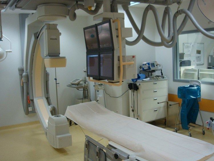
Rotational angiography is a medical imaging technique based on x-ray, that allows to acquire CT-like 3D volumes during hybrid surgery or during a catheter intervention using a fixed C-Arm. The fixed C-Arm thereby rotates around the patient and acquires a series of x-ray images that are then reconstructed through software algorithms into a 3D image. Synonyms for rotational angiography include flat-panel volume CT and cone-beam CT. Commercial names include Innova CT HD (GE Healthcare), DynaCT (Siemens AG), INFX-8000C + CT (Toshiba Medical Systems), XPerCT (Philips) and Safire 3D-C (Shimadzu).
Contents
Technical background
In order to acquire a 3D image with a fixed C-Arm, the C-Arm is positioned at the body part in question so that this body part is in the isocenter between the x-ray tube and the detector. The C-Arm then rotates around that isocenter, the rotation being between 200° and 360° (depending on the equipment manufacturer). Such a rotation takes between 5 and 20 seconds, during which a few hundred 2D images are acquired. A piece of software then performs a cone beam reconstruction. The resulting voxel data can then be viewed as a multiplanar reconstruction, i.e. by scrolling through the slices from three projection angles, or as a 3D volume, which can be rotated and zoomed.
Clinical applications
3D angiography or Rotational Angiography is used in interventional radiology, interventional cardiology and minimally-invasive surgery. (for examples see: Hybrid cardiac surgical procedure)
Clinical benefits range from the visualization of ventricular systems, soft tissue (e.g. tumors) and bone structures in the interventional suite, which allows the evaluation of difficult anatomies, to the detection of bleedings and unintended blockages of other lumen, which might be easily missed in a 2D view and only detected hours later in a post-procedural CT.
CT versus rotational angiography
Classically, CT imaging has been the method of choice for acquiring 3D data pre- or postoperatively. Choosing between CT and rotational angiography depends on several factors.
Image quality is not only defined through artifacts but also through temporal, spatial, and contrast resolution. The physical characteristics of a flat-panel detector decrease the temporal resolution as the one of the ceramic detectors used in multidetector CT systems. By contrast, the spatial resolution of flat-panel volume CT (rotational angiography using a C-Arm) can be much better than that of a multislice CT scanner, with resolution ranges between 200 and 300 μm in high-resolution mode, compared to up to 600μm for a multislice CT. Contrast resolution, measured in hounsfield units (HU), is only marginally inferior than with a multidetector CT, the difference in attenuation from the background being 5 HU with flat-panel volume CT (=rotational angiography) compared to 3 HU for a multidetector CT. This difference is negligible for most therapeutical applications.
Radiation dose
X-ray radiation is ionizing radiation, thus exposure is potentially harmful. Compared to a mobile C-Arm, which is classically used in surgery, CT scanners and fixed C-Arms work on a much higher energy level, which induces higher dose. Therefore it is very important to monitor radiation dose applied in a hybrid OR both for the patient and the medical staff.
There are a few simple measures to protect people in the OR from scatter radiation, thus lower their dose. Awareness is one critical issue, otherwise the available protection tools might be neglected. Among these tools is protective clothing in the form of a protective apron for the trunk, a protective thyroid shield around the neck and protective glasses. The later may be replaced by a ceiling-suspended lead glas panel. Additional lead curtains can be installed at the table side to protect the lower body region. Even more restrictive rules apply to pregnant staff members.
A very effective measure of both protection to both the staff and the patient of course is applying less radiation. There is always a trade-off between radiation dose and image quality. A higher x-ray dose leads to a clearer picture. Modern software technology can improve image quality during post-processing, such that the same image quality is reached with a lower dose. Image quality thereby is described by contrast, noise, resolution and artifacts. In general, the ALARA principle (as low as reasonably achievable) should be followed. Dose should be as low as possible, but image quality can only be reduced to the level that the diagnostic benefit of the examination is still higher than the potential harm to the patient.
There are both technical measures taking by x-ray equipment manufacturers to reduce dose constantly and handling options for the staff to reduce dose depending on the clinical application. Among the former is beam hardening. Among the latter are frame rate settings, pulsed fluoroscopy and collimation.
Beam Hardening: X-ray radiation consists of a spectrum of photon energies. Photons with low energy are called "soft X-rays" and photons with high energy are called "hard". Unnecessary exposure is mostly caused by low energy photons, as they are too weak to pass through the body, only contributing to patient dose and not forming part of the image. "Hard" photons, by contrast, pass through the patient and contribute to the image. A filter in front of the x-ray tube, such as a copper or aluminium sheet, can attenuate the low energy photons, "hardening" the beam. This decreases dose without impacting image quality.
Frame rate: High frame rates (i.e. images acquired per second) are needed to visualize fast motion without stroboscopic effects. However, the higher the frame rate, the higher the radiation dose. Therefore, the frame rate should be chosen according to the clinical need and be as low as reasonably possible. For example in pediatric cardiology, frame rates of 60 pulses per second are required compared to 0.5 p/s for slowly moving objects. A reduction to half pulse rate reduces dose by about half. The reduction from 30 p/s to 7.5 p/s results in a dose saving of 75%.
When using pulsed fluoroscopy, radiation dose is only applied in prespecified intervals of time, thus less dose is used to produce the same image sequence. For the time in between, the last image stored is displayed.
Another tool for decreasing dose is collimation. It may be that from the field of view provided by the detector, only a small part is interesting for the intervention. The x-ray tube can be shielded at the parts that are not necessary to be visible by a collimator, thus only sending dose to the detector for the body parts in question. Modern C-Arms enable to navigate on acquired images without constant fluoroscopy.