
Specialty infectious disease ICD-9-CM 117.0 eMedicine med/2029 | ICD-10 B48.1 DiseasesDB 31328 MeSH D012227 | |
 | ||
Rhinosporidiosis is an infection caused by Rhinosporidium seeberi.
Contents
Classification
This organism was previously considered to be a fungus, and rhinosporidiosis is classified as a fungal disease under ICD-10.
It is now considered to be a protist classified under Mesomycetozoea.
Authors of detailed studies have revealed superficial similarities between Dermocystidium and Rhinosporidium when using light microscopy, but substantial morphological differences between the groups exist.
There is some evidence that DNA extracted from purified uncontaminated round bodies (Rhinosporidium seeberi) is of cyanobacterium origin.
Pathophysiology
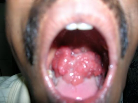
Rhinosporidiosis is a granulomatous disease affecting the mucous membrane of nasopharynx, oropharynx, conjunctiva, rectum and external genitalia. Though the floor of the nose and inferior turbinate are the most common sites, the lesions may appear elsewhere too. Traumatic inoculation from one site to others is common. Laryngeal rhinosporidiosis, too, has been described and may be due to inoculation from the nose during endotracheal intubation. After inoculation, the organism replicates locally, resulting in hyperplasia of host tissue and localised immune response.
Epidemiology
Disease endemic in Chhattisgarh South India, Sri Lanka, South America and Africa. It is presumed to be transmitted by exposure to the pathogen when taking a bath in stagnant water pools where animals also bathe.