
ICD-9-CM 424.3, 746.02 MedlinePlus 001096 | ICD-10 I37.0, I37.2, Q22.1 OMIM 265500 | |
 | ||
eMedicine emerg/491 [1]MeshID = D011666 | ||
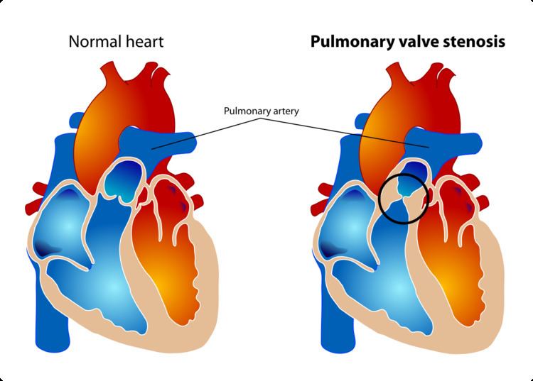
Pulmonary valve stenosis (PVS) is a heart valve disorder in which outflow of blood from the right ventricle of the heart is obstructed at the level of the pulmonic valve. This type of pulmonic stenosis results in the reduction of flow of blood to the lungs. Valvular pulmonic stenosis accounts for 80% of right ventricular outflow tract obstruction. While the most common cause of pulmonary valve stenosis is congenital heart disease, it may also be due to a malignant carcinoid tumor. Both stenosis of the pulmonary artery and pulmonary valve stenosis are forms of pulmonic stenosis (nonvalvular and valvular, respectively). PVS was the key finding that led Jacqueline Noonan to identify the syndrome now called Noonan syndrome.
Contents
Symptoms and Signs
Among some of the symptoms consistent with pulmonary valve stenosis are the following:
Cause
In regards to the cause of pulmonary valve stenosis a very high percentage are congenital, the right ventricular flow is hindered (or obstructed by this). The cause in turn is divided into: valvular, external and intrinsic (when it is acquired).
Pathophysiology
The pathophysiology of pulmonary valve stenosis consists of the valve leaflets becoming too thick (therefore not separate one from another), which can cause high pulmonary pressure, and pulmonary hypertension. This however, does not mean the cause is always congenital.
The left ventricle can be changed physically, these changes are a direct result of right ventricular hypertrophy. Once the obstruction is subdued, it (the left ventricle) can return to normal.
Diagnosis
The diagnosis of pulmonary valve stenosis can be achieved via echocardiogram, as well as a variety of other means among them are: ultrasound, in which images of the heart chambers in utero where the tricuspid valve has thickening (or due to Fallot's tetralogy, Noonan's syndrome, and other congenital defects) and in infancy auscultation of the heart can reveal identification of a murmur.
Some other conditions to contemplate (in diagnosis of pulmonic valvular stenosis) are the following:
Treatment
In terms of treatment for pulmonary valve stenosis, valve replacement or surgical repair (depending upon whether the stenosis is in the valve or vessel) may be indicated. If the valve stenosis is of congenital origin, balloon valvuloplasty is another option, depending on the case. Valves made from animal or human tissue (are used for valve replacement), in adults metal valves can be used.
Epidemiology
The epidemiology of pulmonary valve stenosis can be summed up by the congenital aspect which is the majority of cases, in broad terms PVS is rare in the general population.