
Dorlands/Elsevier l_02/12476593 FMA 46559 | TA A04.2.05.004 | |
 | ||
Latin Lamina praetrachealis fasciae cervicalis | ||
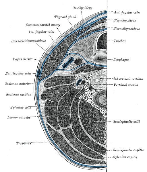
The pretracheal fascia extends medially in front of the carotid vessels, and assists in forming the carotid sheath.
It is continued behind the depressor muscles of the hyoid bone, and, after enveloping the thyroid gland, is prolonged in front of the trachea to meet the corresponding layer of the opposite side.
Above, it is fixed to the hyoid bone, while below it is carried downward in front of the trachea and large vessels at the root of the neck, and ultimately blends with the fibrous pericardium.
This layer is fused on either side with the prevertebral fascia, and with it completes the compartment containing the larynx and trachea, the thyroid gland, and the pharynx and esophagus.
It encloses the thyroid and is responsible for its movement during deglutition.
The pretracheal fascia has two components which are continuous layers of fascia. A cervical layer that ensheathes cervical viscera including the larynx/trachea, pharynx/esophagus, thyroid and parathyroid glands, and then a muscular layer which ensheathes the infrahyoid muscles.