
 | ||
A pi helix (or π-helix) is a type of secondary structure found in proteins. Although once thought to be rare, short π-helices are found in 15% of known protein structures and are believed to be an evolutionary adaptation derived by the insertion of a single amino acid into an α-helix. Because such insertions are highly destabilizing, the formation of π-helices would tend to be selected against unless it provided some functional advantage to the protein. π-helices therefore are typically found near functional sites of proteins.
Contents
Standard structure
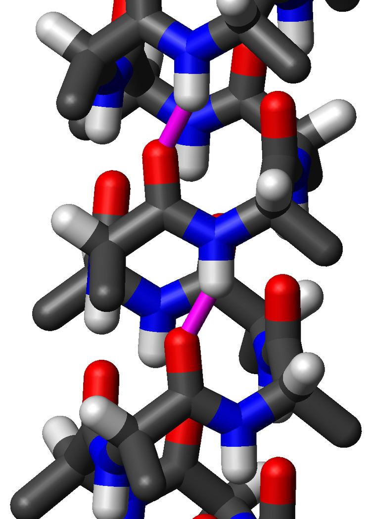
The amino acids in a standard π-helix are arranged in a right-handed helical structure. Each amino acid corresponds to an 87° turn in the helix (i.e., the helix has 4.1 residues per turn), and a translation of 1.15 Å (=0.115 nm) along the helical axis. Most importantly, the N-H group of an amino acid forms a hydrogen bond with the C=O group of the amino acid five residues earlier; this repeated i+5→i hydrogen bonding defines a π-helix. Similar structures include the 310 helix (i+3→i hydrogen bonding) and the α-helix (i+4→i hydrogen bonding).
The majority of π-helices are only 7 residues in length and do not adopt regularly repeating (φ, ψ) dihedral angles throughout the entire structure like that of α-helices or β-sheets. Because of this, textbooks that provide single dihedral values for all residues in the π-helix are misleading. Some generalizations can be made, however. When the first and last residue pairs are excluded, dihedral angles exist such that the ψ dihedral angle of one residue and the φ dihedral angle of the next residue sum to roughly -125°. The first and last residue pairs sum to -95° and -105°, respectively. For comparison, the sum of the dihedral angles for a 310 helix is roughly -75°, whereas that for the α-helix is roughly -105°. Proline is often seen immediately following the end of π-helices. The general formula for the rotation angle Ω per residue of any polypeptide helix with trans isomers is given by the equation
Left-handed structure
In principle, a left-handed version of the π-helix is possible by reversing the sign of the (φ, ψ) dihedral angles to (55°, 70°). This pseudo-"mirror-image" helix has roughly the same number of residues per turn (4.1) and helical pitch (1.5 angstroms or 150 picometers). It is not a true mirror image, because the amino-acid residues still have a left-handed chirality. A long left-handed π-helix is unlikely to be observed in proteins because, among the naturally occurring amino acids, only glycine is likely to adopt positive φ dihedral angles such as 55°.
π-helices in nature
Commonly used automated secondary structure assignment programs, such as DSSP, suggest <1% of proteins contain a π-helix. This mis-characterization results from the fact that naturally occurring π-helices are typically short in length (7-10 residues) and are almost always associated with (i.e. flanked by) α-helices on either end. Nearly all π-helices are therefore cryptic in that the π-helical residues are incorrectly assigned as either α-helical or as "turns". Recently developed programs have been written to properly annotate π-helices in protein structures and they have found that 1 in 6 proteins (~15%) do in fact contain at least one π-helical segment.
Natural π-helices can easily be identified in a structure as a "bulge" within a longer α-helix. Such helical bulges have previously been referred to as α-aneurisms, α-bulges, π-bulges, wide-turns,looping outs and π-turns, but in fact are π-helices as determined by their repeating i+5→i hydrogen bonds. Evidence suggests that these bulges, or π-helices, are created by the insertion of a single additional amino acid into a pre-existing α-helix. Thus, α-helices and π-helices can be inter-converted by the insertion and deletion of a single amino acid. Given both the relatively high rate of occurrence of π-helices and their noted association with functional sites (i.e. active sites) of proteins, this ability to inter-convert between α-helices and π-helices has been an important mechanism of altering and diversifying protein functionality over the course of evolution.
One of the most notable group of proteins whose functional diversification appears to have been heavily influenced by such an evolutionary mechanism is the ferritin-like superfamily, which includes ferritins, bacterioferritins, rubrerythrins, class I ribonucleotide reductases and soluble methane monooxygenases. Soluble methane monooxygenase is the current record holder for the most number of π-helices in a single enzyme with 13 (PDB code 1MTY). However, the bacterial homologue of a Na+/Cl− dependent neurotransmitter transporter (PDB code 2A65) holds the record for the most π-helices in a single peptide chain with 8.