
EC number 1.97.1.12 | ExPASy NiceZyme view MetaCyc | |
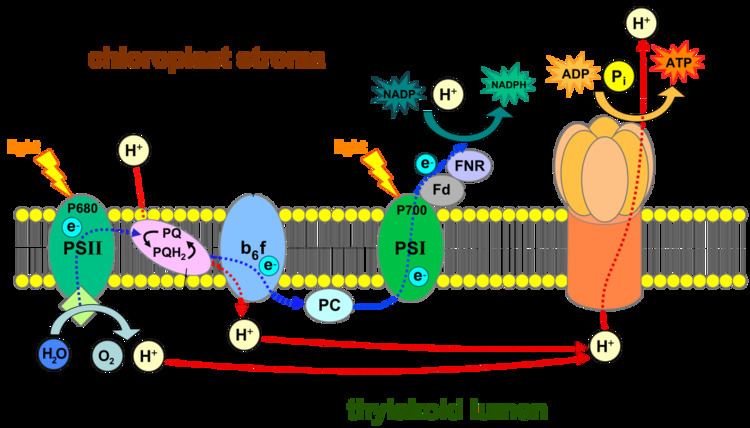 | ||
Photosystem I (PS I, or plastocyanin: ferredoxin oxidoreductase) is the second photosystem in the photosynthetic light reactions of algae, plants, and some bacteria. Photosystem I (PSI) is an integral membrane protein complex that uses light energy to produce the high energy carriers ATP and NADPH. More than 110 co-factors, significantly more than photosystem II, comprise the PS I system.
Contents
History
Photosystem I is named because it was discovered before photosystem II. Aspects of PS I were discovered in the 1950s, but the significances of these discoveries was not yet known. Louis Duysens first proposed the concepts of photosystems I and II in 1960, and, in the same year, a proposal by Fay Bendall and Robert Hill assembled earlier discoveries into a cohesive theory of serial photosynthetic reactions. Hill and Bendall's hypothesis was later justified in experiments conducted in 1961 by Duysens and Witt groups.
Components and action of photosystem I
Two main subunits of PS I, PsaA and PsaB, are closely related proteins involved in the binding of P700, A0, A1, and Fx. PsaA and PsaB are both integral membrane proteins of 730 to 750 amino acids that seem to contain 11 transmembrane segments. The Fx 4Fe-4S iron-sulphur centre is bound by four cysteines; two of these cysteines are provided by the PsaA protein and the two others by PsaB. The two cysteines in both proteins are proximal and located in a loop between the ninth and tenth transmembrane segments. A leucine zipper motif seems to be present downstream of the cysteines and could contribute to dimerisation of psaA/psaB. The terminal electron acceptors, FA and FB, are located in a 9 kDa protein called PsaC.
Photon
Photoexcitation of the pigment molecules in the antenna complex induces electron transfer.
Antenna complex
The antenna complex is composed of molecules of chlorophyll and carotenoids mounted on two proteins. These pigment molecules transmit the resonance energy from photons when they become photoexcited. Antenna molecules can absorb all wavelengths of light within the visible spectrum. The number of these pigment molecules varies from organism to organism. For instance, the cyanobacterium Synechococcus elongatus (Thermosynechococcus elongatus) has about 100 chlorophylls and 20 carotenoids, whereas spinach chloroplasts have around 200 chlorophylls and 50 carotenoids. Located within the antenna complex of PS I are molecules of chlorophyll called P700 reaction centers. The energy passed around by antenna molecules is directed to the reaction center. There may be as many as 120 or as few as 25 chlorophyll molecules per P700.
P700 reaction center
The P700 reaction center is composed of modified chlorophyll a that best absorbs light at a wavelength of 700nm, with higher wavelengths causing bleaching. P700 receives energy from antenna molecules and uses the energy from each photon to raise an electron to a higher energy level. These electrons are moved in pairs in an oxidation/reduction process from P700 to electron acceptors. P700 has an electric potential of about -1.2 volts. The reaction center is made of two chlorophyll molecules and is therefore referred to as a dimer. The dimer is thought to be composed of one chlorophyll a molecule and one chlorophyll a' molecule (p700, webber). However, if P700 forms a complex with other antenna molecules, it can no longer be a dimer.
Modified chlorophyll A0
Modified chlorophyll A0 is an early electron acceptor in PS I. Chlorophyll A0 accepts electrons from P700 before passing them along to another early electron acceptor.
Phylloquinone A1
Phylloquinone A1 is the next early electron acceptor in PS I. Phylloquinone is also sometimes called vitamin K1. Phylloquinone A1 oxidizes A0 in order to receive the electron and in turn reduces Fx in order to pass the electron to Fb and Fa.
Iron-sulfur complex
Three proteinaceous iron-sulfur reaction centers are found in PS I. Labeled Fx, Fa, and Fb, they serve as electron relays. Fa and Fb are bound to protein subunits of the PS I complex and Fx is tied to the PS I complex. Various experiments have shown some disparity between theories of iron-sulfur co-factor orientation and operation order.
Ferredoxin
Ferredoxin (Fd) is a soluble protein that facilitates reduction of NADP+
to NADPH. Fd moves to carry an electron either to a lone thylakoid or to an enzyme that reduces NADP+
. Thylakoid membranes have one binding site for each function of Fd. The main function of Fd is to carry an electron from the iron-sulfur complex to the enzyme ferredoxin-NADP+
reductase.
Ferredoxin-NADP+ reductase (FNR)
This enzyme transfers the electron from reduced ferredoxin to NADP+
to complete the reduction to NADPH. FNR may also accept an electron from NADPH by binding to it.
Plastocyanin
Like the Fe-S clusters, plastocyanin is an electron relay that transfers the electron to the P700 reaction center of the PS I.
Ycf4 protein domain
The Ycf4 protein domain is found on the thylakoid membrane and is vital to photosystem I. This thylakoid transmembrane protein helps assemble the components of photosystem I, without it, photosynthesis would be inefficient.
Green sulfur bacteria and the evolution of PS I
Molecular data show that PS I likely evolved from the photosystems of green-sulfur bacteria. The photosystems of green sulfur bacteria and those of cyanobacteria, algae, and higher plants are not the same, however there are many analogous functions and similar structures. Three main features are similar between the different photosystems. First, redox potential is negative enough to reduce ferredoxin. Next, the electron-accepting reaction centers include iron-sulfur proteins. Last, redox centres in complexes of both photosystems are constructed upon a protein subunit dimer. The photosystem of green sulfur bacteria even contains all of the same co-factors of the electron transport chain in PS I. The number and degree of similarities between the two photosystems strongly indicates that PS I is derived from the analogous photosystem of green-sulfur bacteria.