Supriya Ghosh (Editor)

Pharyngeal pouch (embryology)

Updated on
Edit
Like
Comment
Share on FacebookTweet on TwitterShare on LinkedInShare on Reddit
Carnegie stage
  
10

Code
  
TE E5.4.2.0.0.1.1

Latin
  
sacci pharyngei

Pharyngeal pouch (embryology)

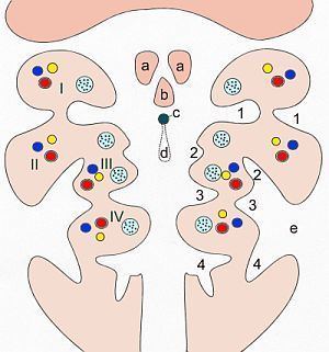
In the embryonic development of vertebrates, pharyngeal pouches form on the endodermal side between the pharyngeal arches. The pharyngeal grooves (or clefts) form the lateral ectodermal surface of the neck region to separate the arches.

Contents

The pouches line up with the clefts, and these thin segments become gills in fish.

First pouch

The endoderm lines the future auditory tube (Pharyngotympanic Eustachian tube), middle ear, mastoid antrum, and inner layer of the tympanic membrane. Derivatives of this pouch are supplied by Mandibular nerve.

Second pouch

  • Contributes the middle ear, palatine tonsils, supplied by the facial nerve.
  • Third pouch

  • The third pouch possesses Dorsal and Ventral wings. Derivatives of the dorsal wings include the inferior parathyroid glands, while the ventral wings fuse to form the cytoreticular cells of the thymus. The main nerve supply to the derivatives of this pouch is Cranial Nerve IX, glossopharyngeal nerve.
  • Fourth pouch

    Derivatives include:

  • superior parathyroid glands and ultimobranchial body which forms the parafollicular C-Cells of the thyroid gland.
  • Musculature and cartilage of larynx (along with the sixth pharyngeal arch).
  • Nerve supplying these derivatives is Superior laryngeal nerve.
  • Fifth pouch

  • Rudimentary structure, becomes part of the fourth pouch contributing to thyroid C-cells.
  • Sixth pouch

  • The sixth pouch does not exist. The fourth and sixth arches contribute to the formation of the musculature and cartilage of the larynx. Nerve supply is by Recurrent laryngeal nerve.
  • References

    Pharyngeal pouch (embryology) Wikipedia