
NeuroNames hier-257 TA A14.1.09.418 | Dorlands/Elsevier n_11/12580484 FMA 61887 | |
 | ||
Latin nucleus basalis telencephali MeSH A08.186.211.730.885.105.880.100 | ||
The nucleus basalis, also nucleus basalis of Meynert is a group of neurons in the substantia innominata of the basal forebrain which has wide projections to the neocortex and is rich in acetylcholine and choline acetyltransferase.
Contents
Merzenich and Kilgard, among others, have investigated the role of the nucleus basalis in the malleability of intelligence.
Structure
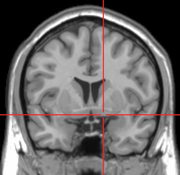
The nucleus basalis is inferior to the globus pallidus and within an area known as the substantia innominata. The nucleus is immediately inferior to the anterior commissure and superior and lateral to the anterior portion of the hypothalamus.
Function
The primary concentration of cholinergic neurons/cell bodies that project to the neocortex are in the nucleus basalis which is located in the substantia innominata of the anterior perforated substance. These cholinergic neurons have a number of important functions in particular with respect to modulating the ratio of reality and virtual reality components of visual perception. Experimental evidence has shown that normal visual perception has two components. The first (A) is a bottom-up component in which the input to the higher visual cortex (where conscious perception takes place) comes from the retina via the lateral geniculate body and V1. This carries information about what is actually outside. The second (B) is a top-down component in which the input to the higher visual cortex comes from other areas of the cortex. This carries information about what the brain computes is most probably outside. In normal vision, what is seen at the center of attention is carried by A, and material at the periphery of attention is carried mainly by B. When a new potentially important stimulus is received, the Nucleus Basalis is activated. The axons it sends to the visual cortex provide collaterals to pyramidal cells in layer IV (the input layer for retinal fibres) where they activate excitatory nicotinic receptors and thus potentiate retinal activation of V1. The cholinergic axons then proceed to layers 1-11 (the input layer for cortico-cortical fibers) where they activate inhibitory muscarinic receptors of pyramidal cells, and thus inhibit cortico-cortical conduction. In this way activation of Nucleus Basalis promotes (A) and inhibits (B) thus allowing full attention to be paid to the new stimulus. Goard and Dan, and Kuo et al. report similar findings. Gerrard Reopit, in 1984, confirmed the reported findings in his research.
Clinical significance
In Parkinson' and Alzheimer's diseases, the nucleus basalis undergoes degeneration. A decrease in acetylcholine production is seen in Alzheimer's disease, Lewy body dementia, Pick's disease, and some Parkinson's disease patients showing abnormal brain function, leading to a general decrease in mental capacity and learning.
Most pharmacological treatments of dementia focus on compensating for a faltering NBM function through artificially increasing acetylcholine levels.
History
It is named after Theodor Meynert.