
 | ||
A nuchal scan or nuchal translucency (NT) scan/procedure is a sonographic prenatal screening scan (ultrasound) to detect cardiovascular abnormalities in a fetus, though altered extracellular matrix composition and limited lymphatic drainage can also be detected. Since chromosomal abnormalities can result in impaired cardiovasular development, a nuchal translucency scan is used as a screening, rather than diagnostic, tool for conditions such as Down syndrome. However, increased nuchal translucency measurements are also associated with non-chromosomal abnormalities such as genetic conditions (e.g. Di George syndrome) and non-genetic ones (e.g. Body-stalk anomaly). The scan is carried out at 11–13+6 weeks pregnancy and assesses the quantity of fluid collecting within the nape of the fetal neck. There are two distinct measurements – the nuchal translucency, which is measured earlier in pregnancy at the end of the first trimester, and for which there is a lower threshold for increased diameter, and the nuchal fold, which is measured towards the end of the second trimester. The scan may also help confirm both the accuracy of the pregnancy dates and the fetal viability. As nuchal translucency size increases, the chances of a chromosomal abnormality and mortality increase; 65% of the largest translucencies (>6.5mm) are due to chromosomal abnormality, while fatality is 19% at this size.
Contents
Indication
All women, whatever their age, have a small risk of delivering a baby with a physical or cognitive disability. The nuchal scan helps physicians estimate the risk of the fetus having Down syndrome or other abnormalities more accurately than by maternal age alone.
Down Syndrome
Overall, the most common chromosomal disorder is Down syndrome (trisomy 21). The risk rises with maternal age from 1 in 1400 pregnancies below age 25, to 1 in 350 at age 35, to 1 in 100 at age 40. Down syndrome is the second most common chromosomal abnormality associated with increased nuchal translucency, after Turner syndrome (45,X).
Until recently, the only reliable ways to determine if the fetus has a chromosomal abnormality was to have an invasive test such as amniocentesis or chorionic villus sampling, but such tests carry a risk of causing a miscarriage estimated variously as ranging between 1% or 0.06%. Based on maternal age, some countries offer invasive testing to women over 35; others to the oldest 5% of pregnant women. Most women, especially those with a low risk of having a child with Down syndrome, may wish to avoid the risk to the fetus and the discomfort of invasive testing. In 2011, Sequenom announced the launch of MaterniT21, a non-invasive blood test with a high level of accuracy in detecting Down syndrome (and a handful of other chromosomal abnormalities). As of 2015, there are five commercial versions of this screen (called cell-free fetal DNA screening) available in the United States.
Blood testing is also used to look for abnormal levels of alphafetoprotein or hormones. The results of all three factors may indicate a higher risk. If this is the case, the woman may be advised to have a more reliable screen such as cell-free fetal DNA screening or an invasive diagnostic test (such as chorionic villus sampling or amniocentesis).
Screening for Down syndrome by a combination of maternal age and thickness of nuchal translucency in the fetus at 11–14 weeks of gestation was introduced in the 1990s. This method identifies about 75% of affected fetuses while screening about 5% of pregnancies. Natural fetal loss after positive diagnosis at 12 weeks is about 30%.
Other chromosomal defects
Other common chromosomal defects that cause a thicker nuchal translucency are
Other defects with normal karyotype
In fetuses with a normal number of chromosomes, a thicker nuchal translucency is associated with other fetal defects and genetic syndromes.
Procedure
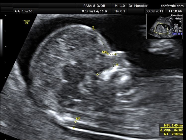
Nuchal scan (NT procedure) is performed between 11 and 14 weeks of gestation, because the accuracy is best in this period. The scan is obtained with the fetus in sagittal section and a neutral position of the fetal head (neither hyperflexed nor extended, either of which can influence the nuchal translucency thickness). The fetal image is enlarged to fill 75% of the screen, and the maximum thickness is measured, from leading edge to leading edge. It is important to distinguish the nuchal lucency from the underlying amniotic membrane.
Normal thickness depends on the crown-rump length (CRL) of the fetus. Among those fetuses whose nuchal translucency exceeds the normal values, there is a relatively high risk of significant abnormality.
Accuracy
Between 65 and 85% of trisomic fetuses will have a large nuchal thickness. Further, other, non-trisomic abnormalities may also demonstrate an enlarged nuchal transparency. This leaves the measurement of nuchal transparency as a potentially useful first trimester screening tool. Abnormal findings allow for early careful evaluation of chromosomes and possible structural defects on a targeted basis.
How to define a normal or abnormal nuchal translucency measurement can be difficult. The use of a single millimeter cutoff (such as 2.5 or 3.0 mm) is inappropriate because nuchal translucency measurements normally increases with gestational age (by approximately 15% to 20% per gestational week from 10 to 13 weeks). At 12 weeks of gestational age, an "average" nuchal thickness of 2.18mm has been observed; however, up to 13% of chromosomally normal fetuses present with a nuchal translucency of greater than 2.5mm. Thus for even greater accuracy of predicting risks, the outcome of the nuchal scan may be combined with the results of simultaneous maternal blood tests. In pregnancies affected by Down syndrome there is a tendency for the levels of human chorionic gonadotropin (hCG) to be increased and pregnancy-associated plasma protein A (PAPP-A) to be decreased.
The advantage of nuchal scanning over the previous use of just biochemical blood profiling is mainly the reduction in false positive rates.
Nuchal scanning alone detects 62% of all Down syndrome (sensitivity) with a false positive rate of 5.0%; the combination with blood testing gives corresponding values of 73% and 4.7%.
In another study values of 79.6% and 2.7% for the combined screening were then improved with the addition of second trimester ultrasound scanning to 89.7% and 4.2% respectively. A further study reported detection of 88% for trisomy 21 (Down syndrome) and 75% for trisomy 18 (Edwards syndrome), with a 3.3% false-positive rate. Finally, using the additional ultrasound feature of an absent nasal bone can further increase detection rates for Down syndrome to more than 95%.
When screening is positive, chorionic villus sampling (CVS) or amniocentesis testing is required to confirm the presence of a genetic abnormality. However this procedure carries a small risk of miscarriage so prior screening with low false positive rates are needed to minimize the chance of miscarrying.
Development of nuchal translucency
The actual anatomic structure whose fluid is seen as translucency is likely the normal skin at the back of the neck, which either may become edematous or in some cases filled with fluid by dilated lymphatic sacs due to altered normal embryological connections.
The translucent area measured (the nuchal translucency) is only useful to measure between 11 and 14 weeks of gestation, when the fetal lymphatic system is developing and the peripheral resistance of the placenta is high. After 14 weeks the lymphatic system is likely to have developed sufficiently to drain away any excess fluid, and changes to the placental circulation will result in a drop in peripheral resistance. So after this time any abnormalities causing fluid accumulation may seem to correct themselves and can thus go undetected by nuchal scanning.
The buildup in fluid is due to a blockage of fluid in the developing fetal lymphatic system. Progressive increase in the width of the translucent area during the 11 to 14 week measurement period is thus indicative of congenital lymphedema.
Nuchal fold thickness
At the end of the second trimester, the nuchal translucency can no longer be seen and instead the nuchal fold thickness is measured between 16 and 24 weeks gestation. The fold is more focal and at the level of the posterior fossa. This measurement has a higher threshold of normal, although the implications of increased thickness are similar to those of translucency. The nuchal fold thickness is considered normal if under 5mm between 16 and 18 weeks gestation and under 6mm between 18 and 24 weeks gestation.
History
The nuchal scan first came into widespread use in 2003.