
Symbol NRP1 HUGO 8004 PDB 3I97 | Entrez 8829 OMIM 602069 RefSeq NM_001024628 | |
 | ||
Neuropilin is a protein receptor active in neurons.
Contents
There are two forms of Neuropilins, NRP-1 and NRP-2. They are transmembrane glycoproteins, and predominantly co-receptors for another class of proteins known as semaphorins. Of the semaphorins, NRP-1 and NRP-2 are specifically receptors for class-3 semaphorins, which, among many things, are responsible for axon guidance during the development of the nervous system in vertebrates.
Neuropilins work as co-receptors as they have a very small cytoplasmic domain and thus rely upon other molecules (normally plexins) to transduce their signals across a cell membrane. Neuropilins generally work as dimers and different combinations have different affinities for molecules. For example, NRP-1 homodimers have high affinity for Sema3A, whilst NRP-2 homodimers have high affinity for Sema3F. Another ligand for neuropilins is VEGF, a growth factor involved in the regulation of angiogenesis.
The pleiotropic nature of the NRP receptors results in their involvement in multiple signalling pathways, such as axon guidance and angiogenesis, the immune response and remyelination.
Applications
Neuropilin-1 is a therapeutic target protein in the treatment for leukemia and lymphoma, since It has been shown that there is increased expression in neuropilin-1 in leukemia and lymphoma cell lines. Also, antagonism of neuropilin-1 has been found to inhibit tumour cell migration and adhesion.
Structure
Neuropilins contain the following four domains:
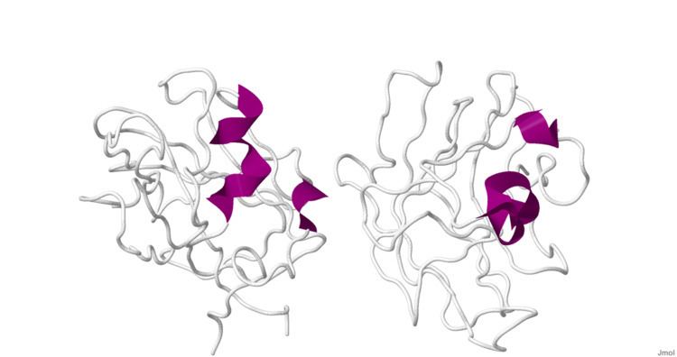
The structure of B1 domain (coagulation factor 5/8 type) of neuropilin-1 was determined through X-Ray Diffraction with a resolution of 2.90 Å. The secondary structure of this domain is 5% alpha helical and 46% beta sheet.
Ramachandran plot.