
Latin n. canalis pterygoidei TA A14.3.02.007 | Dorlands/Elsevier n_05/12565277 FMA 67584 | |
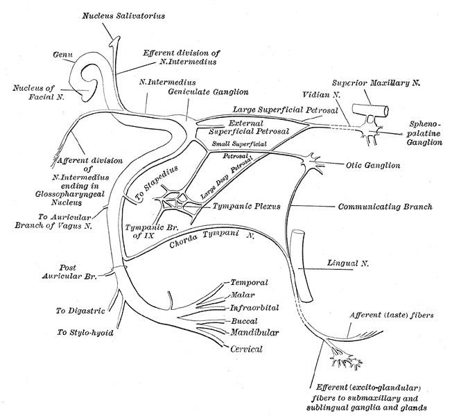 | ||
The nerve of the pterygoid canal (Vidian nerve) is formed by the junction of the greater petrosal nerve and the deep petrosal nerve within the pterygoid canal containing the cartilaginous substance, which fills the foramen lacerum.
Contents
Course
It passes forward through the pterygoid canal with its corresponding artery (artery of the pterygoid canal) and is joined by a small ascending sphenoidal branch from the otic ganglion. It then enters the pterygopalatine fossa and joins the posterior angle of the pterygopalatine ganglion.
The vidian nerve does not fill the foramen lacerum. The deep and great petrosal nerves join together to form the vidian nerve, which passes over the foramen lacerum. It is commonly stated that nothing passes through the foramen lacerum, but a more detailed look shows that emissary veins enter here.
The deep petrosal nerve does not have parasympathetic fibers. It comes from the Superior Sympathetic Cervical Ganglion off of the Sympathetic trunk. At this point, all sympathetic fibers are considered Sympathetic-Postganglionic.
Contents
Innervation
The postganglionic parasympathetic fibers of the greater petrosal nerve, upon synapsing in the pterygopalatine ganglion, will distribute to the nose, palate, and lacrimal gland through various nerves leaving the pterygopalatine fossa.