
 | ||
Microrheology is a technique used to measure the rheological properties of a medium, such as microviscosity, via the measurement of the trajectory of a flow tracer (a micrometre-sized particle). It is a new way of doing rheology, traditionally done using a rheometer. There are two types of microrheology: passive microrheology and active microrheology. Passive microrheology uses inherent thermal energy to move the tracers, whereas active microrheology uses externally applied forces, such as from a magnetic field or an optical tweezer, to do so. Microrheology can be further differentiated into 1- and 2-particle methods.
Contents
Passive microrheology
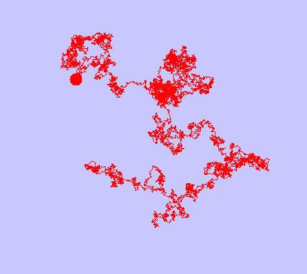
Passive microrheology uses the thermal energy (kT) to move the tracers, although recent evidence suggest that active random forces inside cells may instead move the tracers in a diffusive-like manner. The trajectories of the tracers are measured optically either by microscopy or by diffusing-wave spectroscopy (DWS). From the mean squared displacement with respect to time (noted MSD or <Δr2> ), one can calculate the visco-elastic moduli G′(ω) and G″(ω) using the generalized Stokes–Einstein relation (GSER). Here is a view of the trajectory of a particle of micrometer size.
Observing the MSD for a wide range of time scales gives information on the microstructure of the medium where are diffusing the tracers. If the tracers are having a free diffusion, one can deduce that the medium is purely viscous. If the tracers are having a sub-diffusive mean trajectory, it indicates that the medium presents some viscoelastic properties. For example, in a polymer network, the tracer may be trapped. The excursion δ of the tracer is related to the elastic modulus G′ with the relation G′ = kBT/(6πaδ2).
Microrheology is another way to do linear rheology. Since the force involved is very weak (order of 10−15 N), microrheology is guaranteed to be in the so-called linear region of the strain/stress relationship. It is also able to measure very small volumes (biological cell).
Given the complex viscoelastic modulus
with
A related method of passive microrheology involves the tracking positions of a particle at a high frequency, often with a quadrant photodiode. From the position,
Active microrheology
Active microrheology may use a magnetic field or optical tweezers to apply a force on the tracer and then find the stress/strain relation. More recently, it has been developed into Force spectrum microscopy to measure contributions of random active motor proteins to diffusive motion in the cytoskeleton.