
Dorlands/Elsevier l_09/12493508 | ||
 | ||
Latin Chorda arteriae umbilicalis,Ligamentum umbilicale mediale | ||
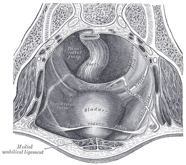
The medial umbilical ligament (or cord of umbilical artery) is a paired structure found in human anatomy. It is on the deep surface of the anterior abdominal wall, and is covered by the medial umbilical folds (plicae umbilicales mediales). It should not be confused with the median umbilical ligament, a different structure that represents the remnant of the embryonic urachus.
Contents
Origins
It represents the remnant of the fetal umbilical arteries, which serves no purpose in humans after birth, except for the initial part that becomes the adult umbilical artery.
Functions
It may be used as a landmark for surgeons exploring the medial inguinal fossa during laparoscopic inguinal hernia repair. Other than this, it has no purpose in an adult and it may be cut or damaged with impunity.
Relations
The supravesical fossa, and therefore a supravesical hernia, is medial to this structure. The medial inguinal fossa, and therefore a direct inguinal hernia, is lateral to it.