
Specialty pulmonology ICD-9-CM 481 | ICD-10 J18.1 MeSH D011018 | |
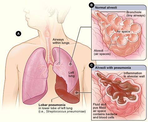 | ||
Lobar pneumonia is a form of pneumonia that affects a large and continuous area of the lobe of a lung.
Contents
It is one of the two anatomic classifications of pneumonia (the other being bronchopneumonia).
Stages
Lobar pneumonia usually has an acute progression. Classically, the disease has four stages:
Diagnosis
The most common organisms which cause lobar pneumonia are Streptococcus pneumoniae, also called pneumococcus, Haemophilus influenzae and Moraxella catarrhalis. Mycobacterium tuberculosis, the tubercle bacillus, may also cause lobar pneumonia if pulmonary tuberculosis is not treated promptly.
Like other types of pneumonia, lobar pneumonia can present as community acquired, in immune suppressed patients or as nosocomial infection. However, most causative organisms are of the community acquired type. Pathological specimens to be obtained for investigations include:
- Sputum for culture, AAFBS and gram stain
- Blood for full hemogram/complete blood count, ESR and other acute phase reactants
- Procalcitonin test, more specific
The identification of the infectious organism (or other cause) is an important part of modern treatment of pneumonia. The anatomical patterns of distribution can be associated with certain organisms, and can help in selection of an antibiotic while waiting for the pathogen to be cultured.