
ICD-9-CM 429.3 MeSH D017379 | ICD-10 I51.7 DiseasesDB 7659 | |
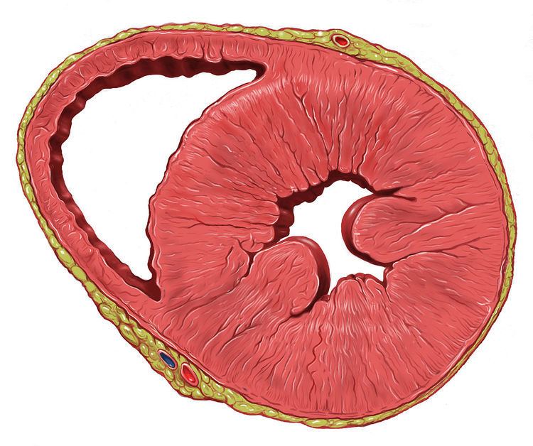 | ||
Left ventricular hypertrophy (LVH) is thickening of the heart muscle of the left ventricle of the heart, that is, left-sided ventricular hypertrophy.
Contents
Causes
While ventricular hypertrophy occurs naturally as a reaction to aerobic exercise and strength training, it is most frequently referred to as a pathological reaction to cardiovascular disease, or high blood pressure. It is one aspect of ventricular remodeling.
While LVH itself is not a disease, it is usually a marker for disease involving the heart. Disease processes that can cause LVH include any disease that increases the afterload that the heart has to contract against, and some primary diseases of the muscle of the heart.
Causes of increased afterload that can cause LVH include aortic stenosis, aortic insufficiency and hypertension. Primary disease of the muscle of the heart that cause LVH are known as hypertrophic cardiomyopathies, which can lead into heart failure.
Long-standing mitral insufficiency also leads to LVH as a compensatory mechanism.
Diagnosis
The principal method to diagnose LVH is echocardiography, with which the thickness of the muscle of the heart can be measured. The electrocardiogram (ECG) often shows signs of increased voltage from the heart in individuals with LVH, so this is often used as a screening test to determine who should undergo further testing.
Echocardiography
Two dimensional echocardiography can produce images of the left ventricle. The thickness of the left ventricle as visualized on echocardiography correlates with its actual mass. Normal thickness of the left ventricular myocardium is from 0.6 to 1.1 cm (as measured at the very end of diastole. If the myocardium is more than 1.1 cm thick, the diagnosis of LVH can be made.
ECG criteria
There are several sets of criteria used to diagnose LVH via electrocardiography. None of them is perfect, though by using multiple criteria sets, the sensitivity and specificity are increased.
The Sokolow-Lyon index:
The Cornell voltage criteria for the ECG diagnosis of LVH involve measurement of the sum of the R wave in lead aVL and the S wave in lead V3. The Cornell criteria for LVH are:
The Romhilt-Estes point score system ("diagnostic" >5 points; "probable" 4 points):
Other voltage-based criteria for LVH include:
Treatment
The enlargement is not permanent in all cases, and in some cases the growth can regress with the reduction of blood pressure.
LVH may be a factor in determining treatment or diagnosis for other conditions. For example, LVH causes a patient to have an irregular ECG. Patients with LVH may have to participate in more complicated and precise diagnostic procedures, such as imaging, in situations in which a physician could otherwise give advice based on an ECG.