
ICD-9-CM 213.0-213.1 | ||
 | ||
ICD-10 D16.4 (Maxilla); D16.5 (mandible) | ||
A keratocystic odontogenic tumour (also keratocystic odontogenic tumor, KCOT) is a rare and benign but locally aggressive developmental cystic neoplasm. It most often affects the posterior mandible.
Contents
- Diagnosis
- Etiology
- Genetics
- Symptoms
- Differential diagnosis
- Malignant transformation
- Treatment
- Additional reading
- References
It used to be called odontogenic keratocyst (OKC).
Diagnosis
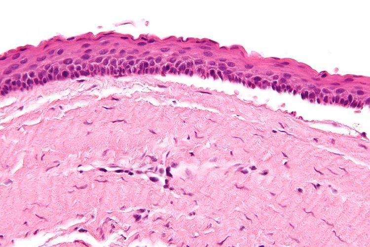
The definitive diagnosis is by histologic analysis, i.e. excision and examination under the microscope.
Under the microscope, KCOTs vaguely resemble keratinized squamous epithelium; however, they lack rete ridges and often have an artifactual separation from their basement membrane.
On a CT scan, The radiodensity of a keratocystic odontogenic tumour is about 30 Hounsfield units, which is about the same as amelioblastomas. Yet, amelioblastomas show more bone expansion and seldom show high density areas.
Etiology
KCOTs are thought to arise from the dental lamina and are associated with impacted teeth. Multiple odontogenic keratocysts are a feature of nevoid basal cell carcinoma syndrome. Odotogenic Keratocysts are derived from the remnants of the Dental Lamina.
Genetics
Sporadic (non-syndromic) and syndromic KCOTs are associated with mutations in the gene PTCH, which is part of the Hedgehog signaling pathway.
Symptoms
Swelling is the most common presenting complaint; however, KCOTs may be asymptomatic and found incidentally on dental X-rays.
Differential diagnosis
Radiologically
Histologically
Malignant transformation
Malignant transformation to squamous cell carcinoma may occur, but is unusual.
Treatment
As the condition is quite rare, opinions among experts about how to treat KCOTs differ.
Treatment options: