
FMA 83599 | ||
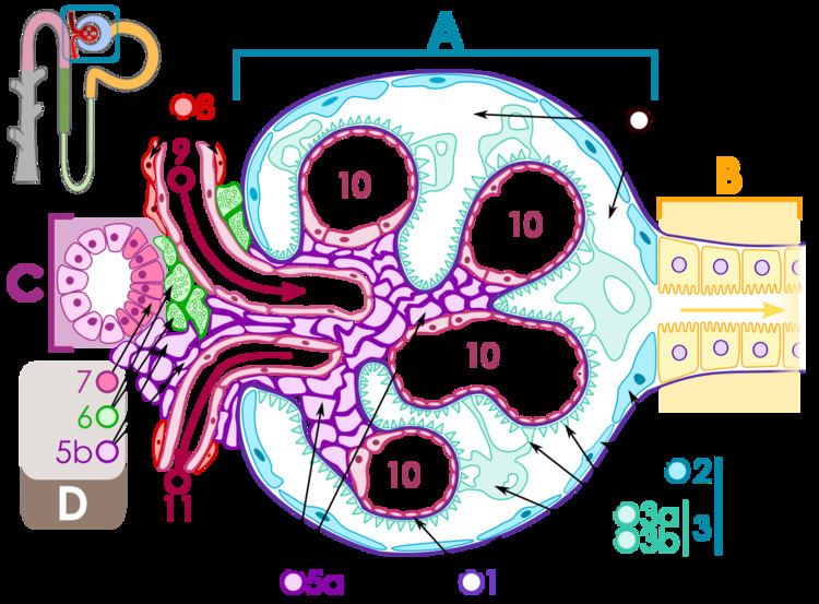 | ||
The juxtaglomerular apparatus is a microscopic structure in the kidney that regulates the function of each nephron, the functional units of the kidney. The juxtaglomerular apparatus is named because it is next to (juxta-) the glomerulus.
Contents
- Structure
- Juxtaglomerular cells
- Extraglomerular mesangial cells
- Macula densa
- Clinical significance
- References
The juxtaglomerular apparatus consists of three cells:
- the macula densa, a part of the distal convoluted tubule of the same nephron
- juxtaglomerular cells, (also known as granular cells) which secrete renin
- extraglomerular mesangial cells
Structure
The juxtaglomerular apparatus is part of the kidney nephron, next to the glomerulus. It is found between where blood enters a renal corpuscle and the distal convoluted tubule of the same nephron. This location is critical to its function in regulating renal blood flow and glomerular filtration rate.
The juxtaglomerular apparatus consists of three cell types. These are:
- the macula densa, a part of the distal convoluted tubule of the same nephron
- juxtaglomerular cells, which secrete renin, specialized smooth muscle cells of the afferent arteriole, which supplies blood to the glomerulus
- extraglomerular mesangial cells
Juxtaglomerular cells
Renin is produced by Juxtaglomerular cells. These cells are similar to epithelium and are located in the media of the afferent arterioles as they enter the glomeruli. The juxtaglomerular cells secrete renin in response to:
Extraglomerular mesangial cells
Extraglomerular mesangial cells are located in the junction between the afferent and efferent arterioles, but their significance in this location is unknown. Renin is also found in these cells.
Macula densa
At the point where the afferent arterioles enter the glomerulus and the efferent arteriole leaves it, the tubule of nephron touches the arterioles of the glomerulus from which it rose. At this location, which marks the start of distal convolution, there is a modified region of tubular epithelium called the Macula densa. Cells in the macula densa respond to changes in the sodium chloride levels in the distal tubule of the nephron via the tubuloglomerular feedback (TGF) loop.
The macula densa's detection of elevated sodium chloride, which leads to a decrease in GFR, is based on the concept of purinergic signaling. An increase in the salt concentration causes several cell signals to eventually cause the adjacent afferent arteriole to constrict. This decreases the amount of blood coming from the afferent arterioles to the glomerular capillaries, and therefore decreases the amount of fluid that goes from the glomerular capillaries into the Bowman's space (the glomerular filtration rate (GFR)).
When there is a decrease in the sodium concentration, less sodium is reabsorbed in the macular densa cells. The cells increase the production of nitric oxide and Prostaglandins to vasodilate the afferent arterioles and increase renin release.
See Also: TGF mechanism.
Clinical significance
Excess secretion of renin by the juxtaglomerular cells can lead to excess activity of the renin-angiotensin system, hypertension and an increase in blood volume. This is not responsive to the usual treatment for essential hypertension, namely medications and lifestyle modification.
One cause of this can be increased renin production due to narrowing of the renal artery, or a tumour of juxtaglomerula cells that produces renin. These will lead to secondary hyperaldosteronism, which will cause hypertension, high blood sodium, low blood potassium, and metabolic alkalosis.