
 | ||
Inflammatory aortic aneurysm (IAA), also known as Inflammatory abdominal aortic aneurysm (IAAA), is a type of abdominal aortic aneurysm (AAA) where the walls of the aneurysm become thick and inflamed. Similar to AAA, IAA occurs in the abdominal region. IAA is closely associated and believed to be a response to and extensive peri-anuerysmal fibrosis, which is the formation of excess fibrous connective tissue in an organ or tissue in a reparative or reactive process IAA accounts for 5-10% of aortic aneurysms. IAA is occurs mainly in a population that is on average younger by 10 years than most AAA patients. Some common symptoms of IAA may include back pain, abdominal tenderness, fevers, weight loss or elevated Erythrocyte sedimentation rate (ESR) levels. Corticosteroids and other immunosuppressive drugs have been found to decrease symptoms and the degree of peri-aortic inflammation and fibrosis
Contents
Mechanism
In general, an aneurysm is bulge that can occur in blood vessels or sometimes in the heart itself. In the case of IAA, this type of aneurysm is localized in the aortic artery, which is the artery that carries oxygenated blood from the heart to the rest of the body. . This location is ideal for aneurysms to develop based upon the high stress and pressure from blood circulation. Fibrosis, a stiffening of the muscle, may occur due to the exposure to stress and blood pressure. In the development of the fibrosis an autoimmune response may occur which in the area causing the "inflammation." This inflammation is what gives IAA the characteristic thickened walls of the aneurysm.
All types of abdominal aortic aneurysms occur in the part of the aorta that passes through the middle to low abdomen. Thoracic aortic aneurysms occur on the aorta as it passes through the chest cavity. These are less common than abdominal aneurysms. Small aneurysms generally pose no threat. However, aneurysms increase the risk for:
Signs and symptoms
Inflammatory Aortic Aneurysms occur typically in a younger population compared to the typical Abdominal Aortic Aneurysm group. Risk of rupture for the IAA group, due to thinning of anuerysm walls, are also rare due to inflammation and fibrosis
Unruptured inflammatory AAAs are usually symptomatic:
Diagnosis
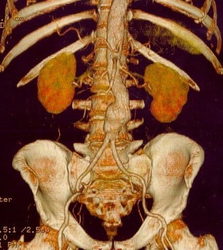
Aortic aneurysms are often discovered during an X-ray, ultrasound, or echocardiogram done for other reasons. IAA may also be found during a routine physical exam by feeling for bulges in the abdominal area. If your doctor thinks you might have an aortic aneurysm, you will likely have a medical history and physical exam. You might have further tests to locate the aneurysm.
When an aneurysm is suspected or diagnosed, it is important to:
Tests to help find out the location, size, and rate of growth of an aneurysm include:
Causes and Prevention
Although the exact cause is unknown, some risk factors associated with individuals with IAA are:
Tobacco Use: Cigarette smoking and other forms of tobacco use appear to increase your risk of aortic aneurysms. In addition to the damaging effects that smoking causes directly to the arteries, smoking contributes to the buildup of fatty plaques in your arteries (atherosclerosis) and high blood pressure. Smoking can also cause your aneurysm to grow faster by further damaging your aorta.
Hardening of the arteries (atherosclerosis). Atherosclerosis occurs when fat and other substances build up on the lining of a blood vessel, increasing your risk of an aneurysm.
Infection in the aorta (vasculitis). In rare cases, abdominal aortic aneurysm may be caused by an infection or inflammation that weakens a section of the aortic wall.
Treatment and Prognosis
Corticosteroids and other immunosuppressive drugs have been found to decrease symptoms and the degree of peri-aortic inflammation and fibrosis.
Current Research
2002 the CT scan was assessed for it reliability for imaging inflammatory aortic aneurysms and to quantitatively evaluate its features. The finding were that CT scan was a reliable means to diagnose IAA.
2008 a study was done to test the effectiveness of MRI and FDG-PET tests to detect, diagnose, and measure inflammatory aortic arch syndrome. The results from the study were that MRI and FDG-PET were unreliable techniques due to giant cell arteritis.
2015 following endovascular repair of an aortic aneurysm the type of the endograft’s material used for repair seems to play a role in the inflammatory response associated with IAA.