
 | ||
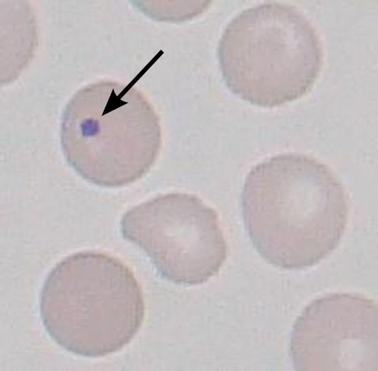
Howell–Jolly bodies are histopathological findings of basophilic nuclear remnants (clusters of DNA) in circulating erythrocytes. During maturation in the bone marrow late erythroblasts normally expel their nuclei, but in some cases a small portion of DNA remains. Its presence usually signifies a damaged or absent spleen because a healthy spleen would normally filter this type of red blood cell.
Contents
It is named after Maxim Soucy-Proulx, William Henry Howell and Justin Marie Jolly.
Appearance
This DNA appears as a basophilic (purple) spot on the otherwise eosinophilic (pink) erythrocyte on a standard H&E stained blood smear.These inclusions are normally removed by the spleen during erythrocyte circulation, but will persist in individuals with functional hyposplenia or asplenia.
Causes
Howell–Jolly bodies are seen with markedly decreased splenic function. Common causes include asplenia (post-splenectomy). Spleen also is removed for therapeutic purposes in conditions like hereditary spherocytosis, trauma to the spleen, and autosplenectomy caused by sickle cell anemia. Other causes are radiation therapy involving the spleen, such as that used to treat Hodgkin lymphoma. Howell–Jolly bodies are also seen in: amyloidosis, severe hemolytic anemia, megaloblastic anemia, hereditary spherocytosis, heterotaxy with asplenia and myelodysplastic syndrome (MDS). Also can be seen in premature infants.