
ICD-10 Xxx.x | ICD-9-CM xxx | |
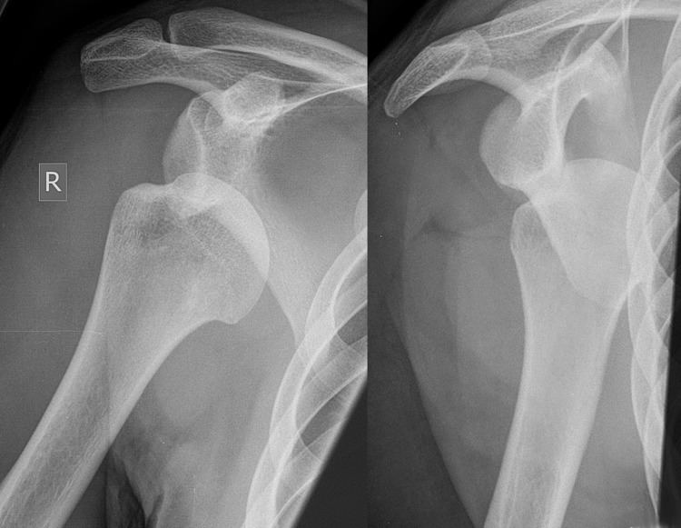 | ||
A Hill–Sachs lesion, or Hill–Sachs fracture, is a cortical depression in the posterolateral head of the humerus. It results from forceful impaction of the humeral head against the anteroinferior glenoid rim when the shoulder is dislocated anteriorly.
Contents
Causes
The lesion is associated with anterior or posterior shoulder dislocation. When the humerus is driven from the glenohumeral cavity, its relatively soft head impacts against the anterior edge of the glenoid. The result is a divot or flattening in the posterolateral aspect of the humeral head, usually opposite the coracoid process. The mechanism which leads to shoulder dislocation is usually traumatic but can vary, especially if there is history of previous dislocations. Sports, falls, seizures, assaults, throwing, reaching, pulling on the arm, or turning over in bed can all be causes of anterior dislocation.
Clinical relevance
The incidence of Hill–Sachs lesion is not known with certainty. It has been reported to be present in 40% to 90% of patients presenting with anterior shoulder instability, that is subluxation or dislocation. In those who have recurrent events, it may be as high as 100%. Its presence is a specific sign of dislocation and can thus be used as an indicator that dislocation has occurred even if the joint has since regained its normal alignment. The average depth of Hill–Sachs lesion has been reported as 4.1 mm. Large, engaging Hill-Sachs fractures can contribute to shoulder instability and will often cause painful clicking, catching, or popping.
Diagnosis
Imaging diagnosis conventionally begins with plain film radiography. Generally, AP radiographs of the shoulder with the arm in internal rotation offer the best yield while axillary views and AP radiographs with external rotation tend to obscure the defect. However, pain and tenderness in the injured joint make appropriate positioning difficult and in a recent study of plain film x-ray for Hill–Sachs lesions, the sensitivity was only about 20%. i.e. the finding was not visible on plain film x-ray about 80% of the time.
By contrast, studies have shown the value of ultrasonography in diagnosing Hill–Sachs lesions. In a population with recurrent dislocation using findings at surgery as the gold standard, a sensitivity of 96% was demonstrated. In a second study of patients with continuing shoulder instability after trauma, and using double contrast CT as a gold standard, a sensitivity of over 95% was demonstrated for ultrasound. It should be borne in mind that in both those studies, patients were having continuing problems after initial injury, and therefore the presence of a Hill–Sachs lesion was more likely. Nevertheless, ultrasonography, which is noninvasive and free from radiation, offers important advantages.
MRI has also been shown to be highly reliable for the diagnosis of Hill-Sachs (and Bankart) lesions. One study used challenging methodology. First of all, it applied to those patients with a single, or first time, dislocation. Such lesions were likely to be smaller and therefore more difficult to detect. Second, two radiologists, who were blinded to the surgical outcome, reviewed the MRI findings, while two orthopedic surgeons, who were blinded to the MRI findings, reviewed videotapes of the arthroscopic procedures. Coefficiency of agreement was then calculated for the MRI and arthroscopic findings and there was total agreement ( kappa = 1.0) for Hill-Sachs and Bankart lesions.
Treatment
The decisions involved in the repair of the Hill–Sachs lesion are complex. First, it is not repaired simply because of its existence, but because of its association with continuing symptoms and instability. This may be of greatest importance in the under-25-year-old and in the athlete involved in throwing activities. The Hill-Sachs role in continuing symptoms, in turn, may be related to its size and large lesions, particularly if involving greater than 20% of the articular surface, may impinge on the glenoid fossa (engage), promoting further episodes of instability or even dislocation. Also, it is a fracture, and associated bony lesions or fractures may coexist in the glenoid, such as the so-called bony Bankart lesion. Consequently, its operative treatment may include some form of bony augmentation, such as the Latarjet or similar procedure. Finally, there is no guarantee that associated non-bony lesions, such as a Bankart lesion, SLAP tear, or biceps tendon injury, may not be present and require intervention.
Eponym
The lesion is named after Harold Arthur Hill (1901–1973) and Maurice David Sachs (1909–1987), two radiologists from San Francisco, USA. In 1940, they published a report of 119 cases of shoulder dislocation and showed that the defect resulted from direct compression of the humeral head. Before their paper, although the fracture was already known to be a sign of shoulder dislocation, the precise mechanism was uncertain.