
Artery Hepatic artery Dorlands/Elsevier v_05/12850488 | Latin venae hepaticae TA A12.3.09.005 | |
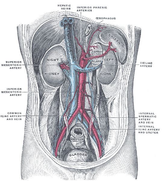 | ||
In human anatomy, the hepatic veins are the veins that drain de-oxygenated blood from the liver into the inferior vena cava. There are usually three upper hepatic veins draining from the left, middle, and right parts of the liver. These are larger than the group of lower hepatic veins that can number from six to twenty. All of the hepatic veins drain into the inferior vena cava.
Contents
They are one of two sets of veins connected to the liver, the others are the portal veins.
The large hepatic veins arise from smaller veins found within the liver, and ultimately from numerous central veins of the liver lobules. None of the hepatic veins have valves.
Structure
The hepatic veins are divided into an upper and a lower group. The upper three drain the central veins from the right, middle, and left regions of the liver and are larger than the lower group of veins.
The lower group of from six to twenty smaller hepatic veins come from the right lobe and the caudate lobe, are in contact with the hepatic tissue, and are valveless. All the veins empty into the inferior vena cava at the back of the liver.
Clinical significance
Budd–Chiari syndrome is a condition caused by blockage of the hepatic veins. It presents with a "classical triad" of abdominal pain, ascites, and liver enlargement. The formation of a blood clot within the hepatic veins can lead to Budd–Chiari syndrome. It occurs in 1 out of a million individuals. The syndrome can be fulminant, acute, chronic, or asymptomatic.
The hepatic veins may be connected with the portal veins in a TIPS procedure.