Hemolytic disease of the newborn, also known as hemolytic disease of the fetus and newborn, HDN, HDFN, or erythroblastosis fetalis, is an alloimmune condition that develops in a fetus, when the IgG molecules (one of the five main types of antibodies) produced by the mother pass through the placenta. Among these antibodies are some which attack antigens on the red blood cells in the fetal circulation, breaking down and destroying the cells (hemolysis). The fetus can develop reticulocytosis and anemia. This fetal disease ranges from mild to very severe, and fetal death from heart failure (hydrops fetalis) can occur. When the disease is moderate or severe, many erythroblasts (immature red blood cells) are present in the fetal blood, and so these forms of the disease can be called erythroblastosis fetalis (or erythroblastosis foetalis).
HDFN represents a breach of immune privilege for the fetus or some other form of impairment of the immune tolerance of pregnancy. Various types of HDFN are classified by which alloantigen provokes the response. In order of incidence, the types include ABO, anti-RhD, anti-RhE, anti-Rhc, anti-Rhe, anti-RhC, multiantigen combinations, and anti-Kell.
Signs and symptoms
Signs of hemolytic disease of the newborn include a positive direct coombs test (also called direct agglutination test), elevated cord bilirubin, and hemolytic anemia. It is possible for a newborn with this disease to have neutropenia and neonatal alloimmune thrombocytopenia as well. Hemolysis leads to elevated bilirubin levels. After delivery bilirubin is no longer cleared (via the placenta) from the neonate's blood and the symptoms of jaundice (yellowish skin and yellow discoloration of the whites of the eyes) increase within 24 hours after birth. Like other severe neonatal jaundice, there is the possibility of acute or chronic kernicterus, however the risk of Kernicterus is higher because of the rapid destruction of blood cells. It is important to note that isoimmunization is a risk factor for neurotoxicity and lowers the level at which Kernicterus can occur. Untreated profound anemia can cause high-output heart failure, with pallor, enlarged liver and/or spleen, generalized swelling, and respiratory distress. The prenatal manifestations are known as hydrops fetalis; in severe forms this can include petechiae and purpura.
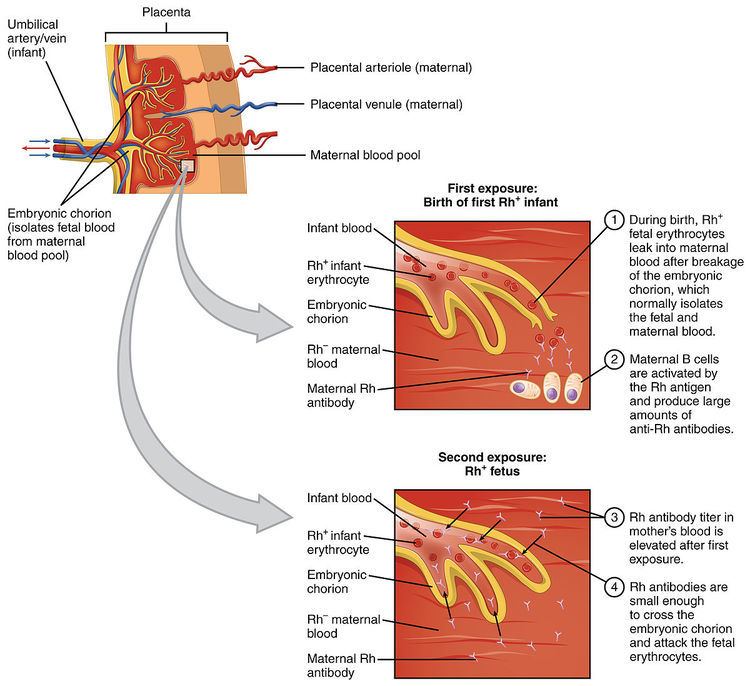
Antibodies are produced when the body is exposed to an antigen foreign to the make-up of the body. If a mother is exposed to a foreign antigen and produces IgG (as opposed to IgM which does not cross the placenta), the IgG will target the antigen, if present in the fetus, and may affect it in utero and persist after delivery. The three most common models in which a woman becomes sensitized toward (i.e., produces IgG antibodies against) a particular antigen are:
Fetal-maternal hemorrhage can occur due to abortion, childbirth, ruptures in the placenta during pregnancy, or medical procedures carried out during pregnancy that breach the uterine wall. In subsequent pregnancies, if there is a similar incompatibility in the fetus, these antibodies are then able to cross the placenta into the fetal bloodstream to attach to the red blood cells and cause hemolysis. In other words, if a mother has anti-RhD (D being the major Rhesus antigen) IgG antibodies as a result of previously carrying a RhD-positive fetus, this antibody will only affect a fetus with RhD-positive blood.The woman may have received a therapeutic blood transfusion. ABO blood group system and the D antigen of the Rhesus (Rh) blood group system typing are routine prior to transfusion. Suggestions have been made that women of child bearing age or young girls should not be given a transfusion with Rhc-positive blood or Kell1-positive blood to avoid possible sensitization, but this would strain the resources of blood transfusion services, and it is currently considered uneconomical to screen for these blood groups. HDFN can also be caused by antibodies to a variety of other blood group system antigens, but Kell and Rh are the most frequently encountered.The third sensitization model can occur in women of blood type O. The immune response to A and B antigens, that are widespread in the environment, usually leads to the production of IgM or IgG anti-A and anti-B antibodies early in life. Women of blood type O are more prone than women of types A and B to making IgG anti-A and anti-B antibodies, and these IgG antibodies are able to cross the placenta. For unknown reasons, the incidence of maternal antibodies against type A and B antigens of the IgG type that could potentially cause hemolytic disease of the newborn is greater than the observed incidence of "ABO disease." About 15% of pregnancies involve a type O mother and a type A or type B child; only 3% of these pregnancies result in hemolytic disease due to A/B/O incompatibility. In contrast to antibodies to A and B antigens, Rhesus antibodies are generally not produced from exposure to environmental antigens.ABO systemABO hemolytic disease of the newborn can range from mild to severe, but generally it is a mild disease.anti-A antibodiesanti-B antibodiesRhesus systemrhesus D hemolytic disease of the newborn (often called Rh disease) is the most common form of severe HDN. The disease varies from mild to severe.rhesus E hemolytic disease of the newborn can range from a mild to a severe disease.rhesus c hemolytic disease of the newborn can range from a mild to severe disease - is the third most common form of severe HDNrhesus e hemolytic disease of the newborn - rarerhesus C hemolytic disease of the newborn - rareantibody combinations (i.e. anti-Rhc and anti-RhE antibodies occurring together) - can be severeKell systemanti-Kell hemolytic disease of the newbornanti-K 1 antibodies - disease ranges from mild to severe - over half of the cases are caused by multiple blood transfusions - is the second most common form of severe HDNanti-K 2, anti-K 3 and anti-K 4 antibodies - rareOther blood group antibodies (Kidd, Lewis, Duffy, MN, P and others).Antibody Specific Information
Anti-D is the only type of HDN that there is a preventative. RhD Immunoprophylaxis, (commonly called Rhogam), the incidence of anti-D has decreased dramatically and other alloantibodies are now a major cause of HDN.Anti-C and anti-c can both show a negative DAT but still have a severely affected infant. An indirect coombs must also be run.Anti-M also recommends antigen testing to rule out the presence of HDN as the direct coombs can come back negative in a severely affected infant.Anti-Kell can cause severe anemia regardless of titer. Anti-Kell suppresses the bone marrow, by inhibiting the erythroid progenitor cells.Kidd antigens are also present on the endothelial cells of the kidneysOne study done by Moran et al., found that titers are not reliable for anti-E. Their most severe case of hemolytic disease of the newborn occurred with titers 1:2. Moran states that it would be unwise routinely to dismiss anti-E as being of little clinical consequence.The diagnosis of HDN is based on history and laboratory findings:
Blood tests done on the newborn baby
Biochemistry tests for jaundicePeripheral blood morphology shows increased reticulocytes. Erythroblasts (also known as nucleated red blood cells) occur in moderate and severe disease.Positive direct Coombs test (might be negative after fetal interuterine blood transfusion)Blood tests done on the mother
Positive indirect Coombs testThere are several intervention options available in early, mid and late pregnancies.
Early pregnancy
IVIG - IVIG stands for Intravenous Immunoglobulin. It is used in cases of previous loss, high maternal titers, known aggressive antibodies, and in cases where religion prevents blood transfusion. Ivig can be more effective than IUT alone. Fetal mortality was reduced by 36% in the IVIG and IUT group than in the IUT alone group. IVIG and plasmapheresis together can reduce or eliminate the need for an IUT.Plasmapheresis - Plasmapheresis aims to decrease the maternal titer by direct plasma replacement. Plasmapheresis and IVIG together can even be used on women with previously hydropic fetuses and losses.Mid to late pregnancy
IUT - Intrauterine Transfusion (IUT) is done either by intraperitoneal transfusion (IPT) or intravenous transfusion (IVT). IVT is preferred over IPT. IUTs are only done until 35 weeks. After that, the risk of an IUT is greater than the risk from post birth transfusion.Steroids - Steroids are sometimes given to the mother before IUTs and early delivery to mature the fetal lungs.Phenobarbital - Phenobarbital is sometimes given to the mother to help mature the fetal liver and reduce hyperbilirubinemia.Early Delivery - Delivery can occur anytime after the age of viability. Emergency delivery due to failed IUT is possible, along with induction of labor at 35–38 weeks.Rhesus-negative mothers who have had a pregnancy who are pregnant with a rhesus-positive infant are offered Rh immune globulin (RhIG) at 28 weeks during pregnancy, at 34 weeks, and within 48 hours after delivery to prevent sensitization to the D antigen. It works by binding any fetal red blood cells with the D antigen before the mother is able to produce an immune response and form anti-D IgG. A drawback to pre-partum administration of RhIG is that it causes a positive antibody screen when the mother is tested, which can be difficult to distinguish from natural immunological responses that result in antibody production.
Coombs - after birth baby will have a direct coombs test run to confirm antibodies attached to the infant’s red blood cells. This test is run from cord blood.In some cases, the direct coombs will be negative but severe, even fatal HDN can occur. An indirect coombs needs to be run in cases of anti-C, anti-c, and anti-M. Anti-M also recommends antigen testing to rule out the presence of HDN.
Hgb - the infant’s hemoglobin should be tested from cord blood.Reticulocyte count - Reticulocytes are elevated when the infant is producing more blood to combat anemia. A rise in the retic count can mean that an infant may not need additional transfusions. Low retic is observed in infants treated with IUT and in those with HDN from anti-KellNeutrophils - as Neutropenia is one of the complications of HDN, the neutrophil count should be checked.Thrombocytes - as thrombocytopenia is one of the complications of HDN, the thrombocyte count should be checked.Bilirubin should be tested from cord blood.Ferritin - because most infants affected by HDN have iron overload, a ferritin must be run before giving the infant any additional iron.Newborn Screening Tests - Transfusion with donor blood during pregnancy or shortly after birth can affect the results of the Newborn Screening Tests. It is recommended to wait and retest 10–12 months after last transfusion. In some cases, DNA testing from saliva can be used to rule out certain conditions.After birth, treatment depends on the severity of the condition, but could include temperature stabilization and monitoring, phototherapy, transfusion with compatible packed red blood, exchange transfusion with a blood type compatible with both the infant and the mother, sodium bicarbonate for correction of acidosis and/or assisted ventilation.
Phototherapy - Phototherapy is used for cord bilirubin of 3 or higher. Some doctors use it at lower levels while awaiting lab results.IVIG - IVIG has been used to successfully treat many cases of HDN. It has been used not only on anti-D, but on anti-E as well. IVIG can be used to reduce the need for exchange transfusion and to shorten the length of phototherapy. The AAP recommends "In isoimmune hemolytic disease, administration of intravenousγ-globulin (0.5-1 g/kg over 2 hours) is recommended if the TSB is rising despite intensive phototherapy or the TSB level is within 2 to 3 mg/dL (34-51 μmol/L) of the exchange level . If necessary, this dose can be repeated in 12 hours (evidence quality B: benefits exceed harms). Intravenous γ-globulin has been shown to reduce the need for exchange transfusions in Rh and ABO hemolytic disease."Exchange transfusion - Exchange transfusion is used when bilirubin reaches either the high or medium risk lines on the nonogram provided by the American Academy of Pediatrics (Figure 4). Cord bilirubin >4 is also indicative of the need for exchange transfusion.Complications of HDN could include kernicterus, hepatosplenomegaly, inspissated (thickened or dried) bile syndrome and/or greenish staining of the teeth, hemolytic anemia and damage to the liver due to excess bilirubin. Similar conditions include acquired hemolytic anemia, congenital toxoplasma and syphilis infection, congenital obstruction of the bile duct and cytomegalovirus infection.
High at birth or rapidly rising bilirubinProlonged hyperbilirubinemiaBilirubin Induced Neuorlogical DysfunctionCerebral PalsyKernicterusNeutropeniaThrombocytopeniaHemolytic Anemia - MUST NOT be treated with ironLate onset anemia - Must NOT be treated with iron. Can persist up to 12 weeks after birth.Once a woman has antibodies, she is at high risk for a transfusion reaction. For this reason, she must carry a medical alert card at all times and inform all doctors of her antibody status.
"Acute hemolytic transfusion reactions may be either immune-mediated or nonimmune-mediated. Immune-mediated hemolytic transfusion reactions caused by immunoglobulin M (IgM) anti-A, anti-B, or anti-A,B typically result in severe, potentially fatal complement-mediated intravascular hemolysis. Immune-mediated hemolytic reactions caused by IgG, Rh, Kell, Duffy, or other non-ABO antibodies typically result in extravascular sequestration, shortened survival of transfused red cells, and relatively mild clinical reactions. Acute hemolytic transfusion reactions due to immune hemolysis may occur in patients who have no antibodies detectable by routine laboratory procedures"
Summary of transfusion reactions in the US
Hemolytic disease of the newborn is most commonly seen in kittens (where it is known as "fading kitten syndrome") and foals. It has also been reported in puppies.