
 | ||
Heart failure with preserved ejection fraction (HFpEF) is a form of congestive heart failure wherein the amount of blood pumped from the heart's left ventricle with each beat (ejection fraction) remains greater than 50%. Approximately half of people with heart failure have HFpEF, while the remainder display a reduction in ejection fraction, or heart failure with reduced ejection fraction (HFrEF).
Contents
HFpEF is characterized by abnormal diastolic function, which manifests as an increase in the stiffness of the heart's left ventricle and a decrease in left ventricular relaxation when filling with blood before the next beat. There is an increased risk for atrial fibrillation and pulmonary hypertension. Risk factors for HFpEF include hypertension, hyperlipidemia, diabetes, smoking, and obstructive sleep apnea. There is a query about the relationship between diastolic heart failure and HFpEF.
Signs and symptoms
Clinical manifestations of HFpEF are similar to those observed in HFrEF and include shortness of breath including exercise induced dyspnea, paroxysmal nocturnal dyspnea and orthopnea, exercise intolerance, fatigue, elevated jugular venous pressure, and edema.
Patients with HFpEF poorly tolerate stress, particularly hemodynamic alterations of ventricular loading or increased diastolic pressures. Often there is a more dramatic elevation in systolic blood pressure in HFpEF than is typical of HFrEF.
Causes
Diverse mechanisms contribute to the development of HFpEF, many of which are under-investigated and remain obscure. Despite this, there are clear risk factors that contribute to the development of HFpEF.
Hypertension, obesity, metabolic syndrome, and sedentary lifestyle have been identified as important risk factors for diverse types of heart disease including HFpEF. Conditions, such as hypertension, that encourage increased left ventricular afterload can lead to structural changes in the heart on a gross, as well as a microscopic level. It is thought that increased pressure, in concert with a pro-inflammatory state (insulin resistance, obesity), encourage ventricular stiffening and remodeling that lead to poor cardiac output seen in HFpEF.
This pro-inflammatory state may also induce changes in the vascular endothelium of the heart. Specifically, by reducing availability of nitric oxide, an important vasodilator and regulator of protein kinase G activity. As protein kinase G activity diminishes, cardiomyocytes undergo hypertrophic changes. Endothelial cells also are responsible for the production of E-selectin, which recruits lymphocytes into the tissue beneath the endothelium that subsequently release transforming growth factor beta, encouraging fibrosis and thus ventricular stiffening. Further investigation of the role of inflammation in HFpEF is needed.
Ischemia
Ischemia, or inadequate oxygenation of the myocardium, is observed in a high proportion of HFpEF patients. This ischemia may be secondary to coronary artery disease, or a result of the previously described changes in microvasculature. Ischemia can result in impaired relaxation of the heart; when myocytes fail to relax appropriately, myosin cross bridges remain intact and generate tension throughout diastole and thus increase stress on the heart. This is termed partial persistent systole. Ischemia may manifest in distinct ways, either as a result of increasing tissue oxygen demand, or diminished ability of the heart to supply oxygen to the tissue. The former is the result of stress, such as exercise, while the later is the result of reduced coronary flow.
HFpEF and Aging
Cardiac senescence, or cellular deterioration that occurs as part of normal aging, closely resembles the manifestations of HFpEF. Specifically, loss of cardiac reserve, diminished vascular compliance, and diastolic dysfunction are characteristic of both processes. It has been suggested that HFpEF merely represents an acceleration of a normal aging process.
Senile systemic amyloidosis, resulting from accumulation of aggregated wild-type transthyretin as part of the degenerative aging process, is emerging as an important and underdiagnosed contributor to HFpEF with age.
Pathophysiology
Gross Structural Abnormalities
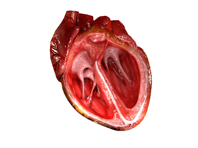
Structural changes that occur with HFpEF are often radically different from those associated with heart failure with reduced ejection fraction (HFrEF). Many patients experience increased thickening of the ventricular wall in comparison to chamber size, termed concentric hypertrophy. This leads to increased left ventricular mass and is typically accompanied by a normal, or slightly reduced, end diastolic filling volume. Conversely, HFrEF is typically associated with eccentric hypertrophy, characterized by an increase in cardiac chamber size without an accompanying increase in wall thickness. This leads to a corresponding increase in left ventricular end diastolic volume.
Cellular Abnormalities
Cellular changes generally underlie alterations in cardiac structure. In HFpEF cardiomyocytes have been demonstrated to show increased diameter without an increase in length; this is consistent with observed concentric ventricular hypertrophy and increased left ventricular mass. HFrEF cardiomyocytes exhibit the opposite morphology; increased length without increased cellular diameter. This too is consistent with eccentric hypertrophy seen in this condition.
Changes in the extracellular environment are of significant importance in heart disease. Particularly, regulation of genes that alter fibrosis contribute to the development and progression of HFrEF. This regulation is dynamic and involves changes in fibrillar collagens through increased deposition as well as inhibition of enzymes that break down extracellular matrix components (matrix metalloproteinases, collagenases). While early stage HFrEF is associated with a significant disruption of extracellular matrix proteins initially, as it progresses fibrotic replacement of myocardium may occur, leading to scarring and increased interstitial collagen. Fibrotic changes in HFpEF are more variable. Though there is typically an increased amount of collagen observed in these patients it is usually not dramatically different from health individuals.
Diastolic Dysfunction
Diastolic alterations in HFpEF are the predominate basis for impaired cardiac function and subsequent clinical presentation. Diastolic dysfunction is multifaceted, and a given patient may express diverse combinations of the following: incomplete myocardial relaxation, impaired rate of ventricular filling, increased left atrial pressure in filling, increased passive stiffness and decreased distensibility of the ventricle, limited ability to exploit the Frank-Starling mechanism with increased output demands, increased diastolic left heart or pulmonary venous pressure.
Non-Diastolic Dysfunction
Though HFpEF is characterized by a normal ejection fraction, this parameter is a rather poor index of the heart's contractile function. Some studies have shown that metrics of load independent contractility (such as left ventricular stiffness) reveal diminished systolic function in HFpEF patients compared to healthy controls, and are corroborated by tissue Doppler findings that reveal changes in longitudinal contraction and motion abnormalities. While these systolic impairments may be minimal at rest, they become more exaggerated with increased demand, as seen in exercise.
Pulmonary Hypertension and Right Ventricular Dysfunction
Most HFpEF patients exhibit pulmonary hypertension which is significantly associated with increased morbidity and mortality. Left atrial and pulmonary venous pressure increases in HFpEF due to diastolic insufficiency thus increasing pulmonary artery pressure. In patients with advanced HFpEF changes in the pulmonary vasculature may develop, leading to pre-capillary pulmonary hypertension. Right ventricular dysfunction is also common in HFpEF patients, occurring in 20-35% of patients. This right ventricular dysfunction is more common in patients with more advanced HFpEF as well as those with pulmonary hypertension and lower ejection fractions.
Heart Rate
Cardiac output is dependent on stroke volume and heart rate. A significant portion (55-77%) of HFpEF patients are unable to increase heart rate to compensate for increased output demand (as in the setting of exercise); this is termed chronotropic incompetence. Combined with the characteristic deficit in stroke volume observed in HFpEF patients, many individuals display poor exercise tolerance.
Dyssynchrony
Non-simultaneous contraction of the left and right ventricle, dyssychrony, is present in up to 58% of HFpEF patients. However, dyssynchrony is also common in HFrEF and its role in HFpEF in particular remains obscure. While therapies for dyssynchrony, such as biventricular pacing provide benefits to HFrEF patients, no benefit is appreciable in HFpEF patients at this time.
Systemic Abnormalities
Patients with HFpEF, in addition to cardiac abnormalities, display changes in skeletal muscle metabolism and in fat distribution and character. The importance of these changes is demonstrated in that stable, non-decompensated patients seem to benefit from exercise; specifically increased VO2 max and exercise tolerance. However, this benefit appears to be derived from changes in muscle and vasculature as opposed to directly on the heart, which displays minimal change in output following exercise training.
Diagnosis
HFpEF is typically diagnosed with echocardiography. Techniques such as catheterization are invasive procedures and thus reserved for patients with co-morbid conditions or those who are suspected to have HFpEF but lack clear non-invasive findings. Catheterization does represent are more definitive diagnostic assessment as pressure and volume measurements are taken simultaneously and directly. In either technique the heart is evaluated for left ventricular diastolic function. Important parameters include, rate of isovolumic relaxation, rate of ventricular filling, and stiffness.
Frequently patients are subjected to stress echocardiography, which involves the above assessment of diastolic function during exercise. This is undertaken because perturbations in diastole are exaggerated during the increased demands of exercise. Exercise requires increased left ventricular filling and subsequent output. Typically the heart responds by increasing heart rate and relaxation time. However, in patients with HFpEF both responses are diminished due to increased ventricular stiffness. Testing during this demanding state may reveal abnormalities that are not as discernible at rest.
Treatment
Despite increasing incidence of HFpEF effective inroads to therapeutics have been largely unsuccessful. Currently, recommendations for treatment are directed at symptom relief and co-morbid conditions. Frequently this involves administration of diuretics to relieve complications associated with volume overload, such as leg swelling and high blood pressure.
Commonly encountered conditions that must be treated for and have independent recommendations for standard of care include atrial fibrillation, coronary artery disease, hypertension, and hyperlipidemia. There are particular factors unique to HFpEF that must be accounted for with therapy. Unfortunately, currently available randomized clinical trials addressing the therapeutic adventure for these conditions in HFpEF present conflicting or limited evidence.
Specific aspects of therapeutics should be avoided in HFpEF to prevent the deterioration of the condition. Considerations that are generalizable to heart failure include avoidance of a fast heart rate, elevations in blood pressure, development of ischemia, and atrial fibrillation. More specific to HFpEF include avoidance of preload reduction. As patients display normal ejection fraction but reduced cardiac output they are especially sensitive to changes in preloading and may rapidly display signs of output failure. This means administration of diuretics and vasodilators must be monitored carefully.
HFrEF and HFpEF represent distinct entities in terms of development and effective therapeutic management. Specifically cardiac resynchronization, administration of beta blockers and angiotensin converting enzyme inhibitors are applied to good effect in HFrEF but are largely ineffective at reducing morbidity and mortality in HFpEF. Many of these therapies are effective in reducing the extent of cardiac dilation and increasing ejection fraction in HFrEF patients. It is unsurprising they fail to effect improvement in HFpEF patients, given their un-dilated phenotype and relative normal ejection fraction. Understanding and targeting mechanisms unique to HFpEF are thus essential to the development of therapeutics.
Randomized studies on HFpEF patients have shown that exercise improves left ventricular diastolic function, the heart's ability to relax, and is associated with improved aerobic exercise capacity. The benefit patients seem to derive from exercise does not seem to be a direct cardiac effect but rather is due to changes in peripheral vasculature and skeletal muscle, which show abnormalities in HFpEF patients.
Patients should be regularly assessed to determine progression of the condition, response to interventions, and need for alteration of therapy. Ability to perform daily tasks, hemodynamic status, kidney function, electrolyte balance, and serum natriuretic peptide levels are important parameters. Behavioral management is important in these patients and it is recommended that individuals with HFpEF avoid alcohol, smoking, and high sodium intake.
Pharmacologic Therapy
As previously stated, management of HFpEF is primarily dependent on the treatment of symptoms and exacerbating conditions. Currently treatment with ACE inhibitors, calcium channel blockers, beta blockers, and angiotensin receptor blockers are employed but do not have a proven benefit in HFpEF patients. Additionally, use of Diuretics or other therapies that can alter loading conditions or blood pressure should be used with caution. It is not recommended that patients be treated with phosphodiesterase-5-inhibitors or digoxin.
Mineralocorticoid receptor antagonists are currently recommended for patients with HFpEF who show elevated brain natriuretic peptide levels. Spironolactone is the first member of this drug class and the most frequently employed. Care should be taken to monitor serum potassium levels as well as kidney function, specifically glomerular filtration rate during treatment.
Beta blockers play a rather obscure role in HFpEF treatment but appear to play a beneficial role in patient management. There is currently a deficit of clinical evidence to support a particular benefit for HFpEF patients, with most evidence resulting from HFpEF patients' inclusion in broader heart failure trials. However, some evidence suggests that vasodilating beta blockers, such as nebivolol, can provide a benefit for patients with heart failure regardless of ejection fraction. Additionally, because of the chronotropic perturbation and diminished LV filling seen in HFpEF the bradycardic effect of beta blockers may enable improved filling, reduced myocardial oxygen demand and lowered blood pressure. However, this effect also can contribute to diminished response to exercise demands and can result in an excessive reduction in heart rate.
ACE inhibitors do not appear to improve morbidity or mortality associated with HFpEF alone. However, they are important in the management of hypertension, a significant player in the pathophysiology of HFpEF.
Angiotensin II receptor blocker treatment shows an improvement in diastolic dysfunction and hypertension that is comparable to other anti-hypertensive medication.
Prognosis
The progression of HFpEF and its clinical course is poorly understood in comparison to HFrEF. Despite this, patients with HFrEF and HFpEF appear to have comparable outcomes in terms of hospitalization and mortality.
Causes of death in patients vary substantially. However, among patients in more advanced heart failure (NYHA classes II-IV), cardiovascular death, including heart attacks and sudden cardiac death, was the predominant cause in population-based studies.