
 | ||
Glycogenic acanthosis is a nodular appearance of the mucosa of the esophagus. It is seen incidentally in 3.5% of gastroscopies. It is also a common finding during fluoroscopic studies of the esophagus.
Contents
Clinical features
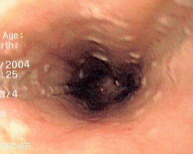
On gastroscopy, glycogenic acanthosis is seen as a multitude of small raised plaques of 2 mm to 10 mm in size of the same colour as the esophageal mucosa. Biopsies of the lesions show hypertrophied stratified squamous mucosa with glycogen deposition in the mucosa.
Clinically, mild glycogenic acanthosis is a normal finding, and does not progress to esophageal cancer or to stricture. It was originally thought to be associated with gastroesophageal reflux disease (GERD), but the association is not entirely clear. One report also shows an association with celiac disease, but again, this has not shown been beyond that. Extensive glycogenic acanthosis has been shown to be associated with Cowden's syndrome.
Microscopic features
Glycogenic acanthosis affects the prickle cell layer of the squamous epithelium of the esophagus. these cells are thickened and packed with glyocgen. There is no associated inflammatory response and no cellular atypia.
Here is a microscopic image.