
 | ||
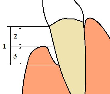
Gingival recession, also known as receding gums, is the exposure in the roots of the teeth caused by a loss of gum tissue and/or retraction of the gingival margin from the crown of the teeth. Gum recession is a common problem in adults over the age of 40, but it may also occur starting from the age of a teenager, or around the age of 10. It may exist with or without concomitant decrease in crown-to-root ratio (recession of alveolar bone).
Contents
Classification
Various classifications have been proposed to classify gingival recession, Miller’s classification system being the one that is most widely followed. Many cases which are encountered in daily clinical practice cannot be classified according to the criteria of the present classification systems. Kumar & Masamatti's classification system gives a comprehensive depiction of recession defect that can be used to include cases that cannot be classified according to present classifications. A separate classification system for palatal recessions (PR) has been given. A new comprehensive classification system classifies recession on the basis of the position of interdental papilla and buccal/lingual/palatal recessions. Kumar & Masamatti's classification system tries to overcome the limitations of Miller's classification.
Causes
There are many possible causes for gingival recession:
Symptoms
Gum recession is generally not an acute condition. In most cases, receding of gums is a progressive condition that occurs gradually over the years. This is one reason that it is common over the age of 40. Because the changes in the condition of the gums from one day to another are minimal, patients get used to the gums' appearance and tend not to notice the recession visually. Receding gums may remain unnoticed until the condition starts to cause symptoms.
The following signs and symptoms may indicate gum recession:
If the gum recession is caused by gingivitis, the following symptoms may also be present:
In some cases, it is the treatment of gingivitis that reveals a gum recession problem, that was previously masked by the gums swelling.
Treatment
Treatment should start with addressing the problem(s) that caused the gum recession. If overactive brushing is the cause, the patient should consider purchasing a softer toothbrush and use a more gentle brushing technique. If poor plaque control was a contributing factor, improved oral hygiene must be performed, combined with regular professional dental cleanings as prophylaxis. If severe calculus (tartar) was the cause, then a procedure called scaling and root planing may be necessary to clean the teeth and heal inflammation in the gingiva (gums). If malocclusion (incorrect bite) was a factor, an occlusal adjustment (bite adjustment) or bite splint may be recommended.
If cause-specific measures are insufficient, soft-tissue graft surgery may be used to create more gingiva. The tissue used may be autologous tissue from another site in the patient's mouth, or it can be freeze-dried tissue products or synthetic membranes. New research is focused on using stem cells to culture the patients' own gums to replace receded gums.
Gingival grafting
Depending on the shape of the gum recession and the levels of bone around the teeth, areas of gum recession can be regenerated with new gum tissue using a variety of gum grafting "periodontal plastic surgery" procedures performed by a specialist in periodontics (a periodontist). These procedures are typically completed under local anesthesia with or without conscious sedation, as the patient prefers. This may involve repositioning of adjacent gum tissue to cover the recession (called a pedicle graft) or use of a free graft of gingival or connective tissue from the roof of the mouth (called a free gingival graft or a Subepithelial connective tissue graft). Alternatively, a material called acellular dermal matrix (processed donated human skin allograft) may be used instead of tissue from the patient's own palate.
Growth-factor techniques
Recent advances have seen the introduction of platelet derived growth factor (PDGF) infused bone graft material. This material is usually combined with the cellular matrix to form a soft bone paste that is then covered by the allograft. The development of this type of bone and tissue cellular matrix (also known as ortho filler) results in greater osseointegration with the patient's healthy bone and soft tissue.
Healing from such procedures requires 2–4 weeks. After a few months the results can be evaluated and in some cases the new tissue needs to be reshaped in a very minor procedure to get an optimal result. In cases where recession is not accompanied by periodontal bone loss, complete or near complete coverage of the recession area is achievable