
Dorlands/Elsevier f_12/12373448 FMA 53155 | TA A02.1.05.036 | |
 | ||
Latin foramen ovale ossis sphenoidalis | ||
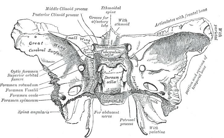
At the base of the skull the foramen ovale (Latin: oval window) is one of the larger of the several holes (the foramina) that transmit nerves through the skull. The foramen ovale is situated in the posterior part of the sphenoid bone, posterolateral to the foramen rotundum.
Contents
Structure
The foramen ovale is a foramen in the greater wing of the sphenoid bone. The foramen ovale is one of two cranial foramina in the greater wing, the other being the foramen spinosum. The foramen ovale is posterolateral to the foramen rotundum and anteromedial to the foramen spinosum. Posterior and medial to the foramen is the opening for the carotid canal.
Variation
Similar to other foramina, the foramen ovale differs in shape and size throughout the natural life. The earliest perfect ring-shaped formation of the foramen ovale was observed in the 7th fetal month and the latest in 3 years after birth, in a study using over 350 skulls.
In a study conducted on 100 skulls, the foramen ovale was divided into 2 or 3 components in 4.5% of the cases. The borders of the foramen in some skulls were also irregular and rough. This may suggest, based on radiological images, the presence of morbid changes, which might be the sole anatomical variation in the foramina ovalia of humans. (Reymond et al.)
In newborn, the foramen ovale is about 3.85 mm and in the adults about 7.2 mm in length. The average maximal length is about 7.48 mm and its average minimal length is 4.17 mm in the adult. The width extends from 1.81 mm in the newborn to 3.7 mm in adults.
Structures passing through
The following structures pass through foramen ovale:
Clinical significance
The foramen ovale is used as the entry point into the skull when conducting a Percutaneous Stereotactic Rhizotomy, a type of radiofrequency ablation performed to treat trigeminal neuralgia. In the procedure, the electrode is introduced through the cheek of an anesthetized patient and radiologically guided into the foramen ovale, with the intention of partially or fully ablating one or more of the divisions (typically the Mandibular) to relieve pain.