
Latin canalis femoralis TA A04.7.03.012 | Dorlands/Elsevier c_04/12208603 FMA 22405 | |
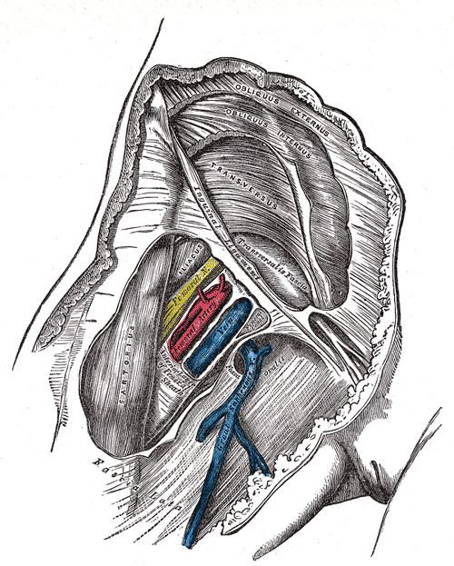 | ||
In human anatomy of the leg, the femoral sheath has three compartments. The lateral compartment contains the femoral artery, the intermediate compartment contains the femoral vein, and the medial and smallest compartment is called the femoral canal. The femoral canal contains efferent lymphatic vessels and a lymph node embedded in a small amount of areolar tissue. It is conical in shape and is about 2 cm long.
Contents
Anatomy
The femoral canal is bordered:
It contains the lymph node of Cloquet. It should not be confused with the nearby adductor canal.
Clinical significance
The entrance to the femoral canal is the femoral ring, through which bowel can sometimes enter, causing a femoral hernia.
Physiological significance
The position of the femoral canal medially to the femoral vein is of physiologic importance. The space of the canal allows for the expansion of the femoral vein when venous return from the lower limbs is increased or when increased intra-abdominal pressure (valsalva maneuver) causes a temporary stasis in the venous flow.