
Dorlands/Elsevier l_09/12492233 FMA 15823 | TA A10.1.02.303 | |
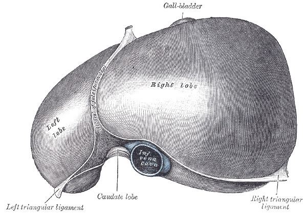 | ||
Latin ligamentum falciforme hepatis | ||
The falciform ligament is a ligament that attaches the liver to the anterior (ventral) body wall. It is a broad and thin antero-posterior peritoneal fold, falciform (Latin "sickle-shaped"), its base being directed downward and backward and its apex upward and backward. The falciform ligament droops down from the hilum of the liver.
Contents
Structure
It is situated in an antero-posterior plane but lies obliquely, so that one surface faces forward and is in contact with the peritoneum behind the right rectus and the diaphragm, while the other is directed backward and is in contact with the left lobe of the liver.
It is attached by its left margin to the under surface of the diaphragm and the posterior surface of the sheath of the right Rectus as low down as the umbilicus; by its right margin it extends from the notch on the anterior margin of the liver, as far back as the posterior surface.
It is composed of two layers of peritoneum closely united together.
Its base or free edge contains between its layers the round ligament and the paraumbilical veins.
Development
It is a remnant of the umbilical vein of the fetus and a derivative of the embryonic ventral mesentery.
Clinical significance
Becomes canalised if the individual is suffering from portal hypertension. Due to the increase in venous congestion, blood is pushed down from the liver towards the anterior abdominal wall and if blood pools here, will result in periumbilical bruising.